

Mutation and immune profiling of metaplastic breast cancer: Correlation with survival
- PMID: 31693690
- PMCID: PMC6834262
- DOI: 10.1371/journal.pone.0224726
Mutation and immune profiling of metaplastic breast cancer: Correlation with survival
Abstract
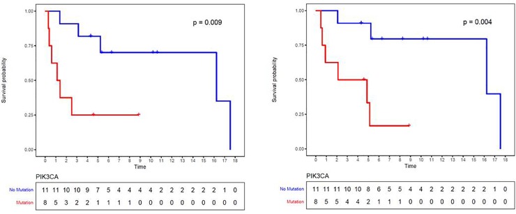
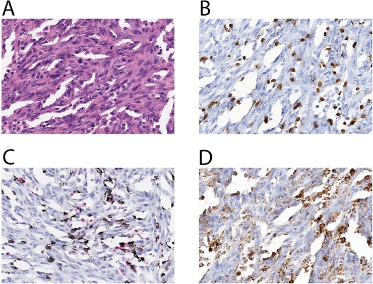
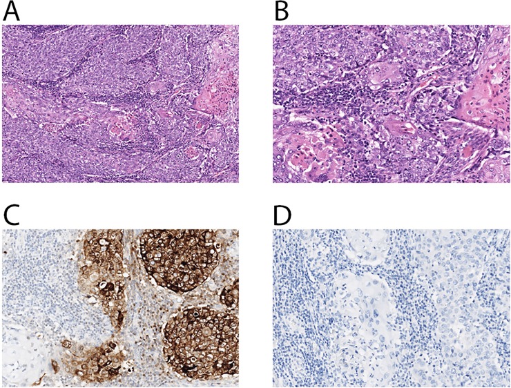
The goal of this study is to characterize the genomic and immune profiles of metaplastic breast cancer (MpBC) and identify the association with survival through an analysis of archived tumor tissue. A next-generation sequencing-based mutational assay (Onco-48) was performed for 21 MpBC patients. Clinicopathologic characteristics were captured, including relapse free survival (RFS) and overall survival (OS). Immunohistochemistry (IHC) for CD3, CD4, CD8, and programmed death-ligand 1 (PD-L1) was also performed. Recurrence free survival (RFS) at 5 years was 57% (95% CI 0.34-0.75) and overall survival (OS) at 5 years was 66% (95% CI 0.41-0.82). The most commonly altered genes were TP53 (68.4%, 13/19), PIK3CA (42.1%, 8/19), and PTEN (15.8%, 3/19. For patients with PIK3CA mutations, RFS and OS were significantly worse than for those without (HR 5.6, 95% CI 1.33-23.1 and HR 8.0, 95% CI 1.53-41.7, respectively). Cox regression estimated that PD-L1 expression was associated with worse RFS and OS (HR 1.08, 95% CI 1.01-1.16 and HR 1.05, 95% CI 1.00-1.11, respectively, for an absolute increase in PD-L1 expression of 1%). In conclusion, PIK3CA mutation and PD-L1 expression confer poor prognosis in this cohort of patients with MpBC.
Conflict of interest statement
YY has contracted clinical trials and research projects sponsored by Merck, Eisai, Novartis, Genentech, and Pfizer, independent of the study presented in this manuscript. There are no patents, products in development or marketed products associated with this research to declare. This does not alter the authors' adherence to PLOS ONE policies on sharing data and materials. The other authors declare that they have no competing interests.
Figures


References
-
- Tavassoli FA. Classification of metaplastic carcinomas of the breast. Pathology annual. 1992;27 Pt 2:89–119. - PubMed
-
- Kurman RJ, Carcangiu ML, Herrington S, Young RH. WHO classification of tumours of female reproductive organs: IARC; 2014.
Publication types
MeSH terms
Substances
Grants and funding
LinkOut - more resources
Full Text Sources
Medical
Research Materials
Miscellaneous
