Review
. 2020 Jan;287(2):222-238.
doi: 10.1111/febs.15127. Epub 2019 Nov 21.

Phosphoinositides in autophagy: current roles and future insights

Affiliations
Review

Phosphoinositides in autophagy: current roles and future insights

Lavinia Palamiuc et al. FEBS J. 2020 Jan.

Abstract

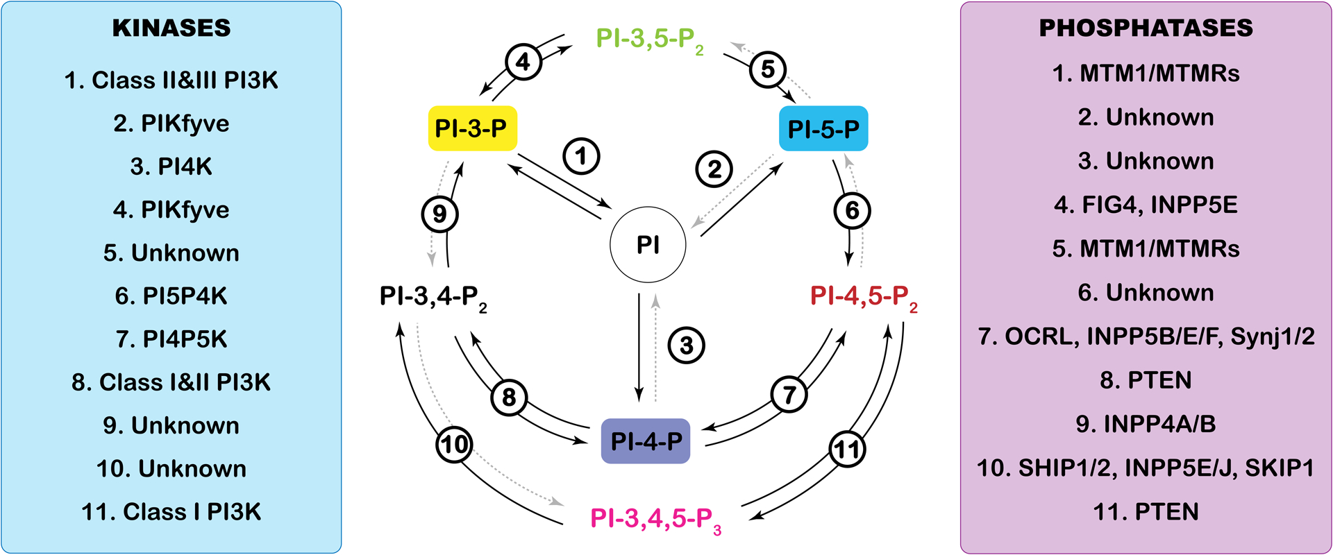
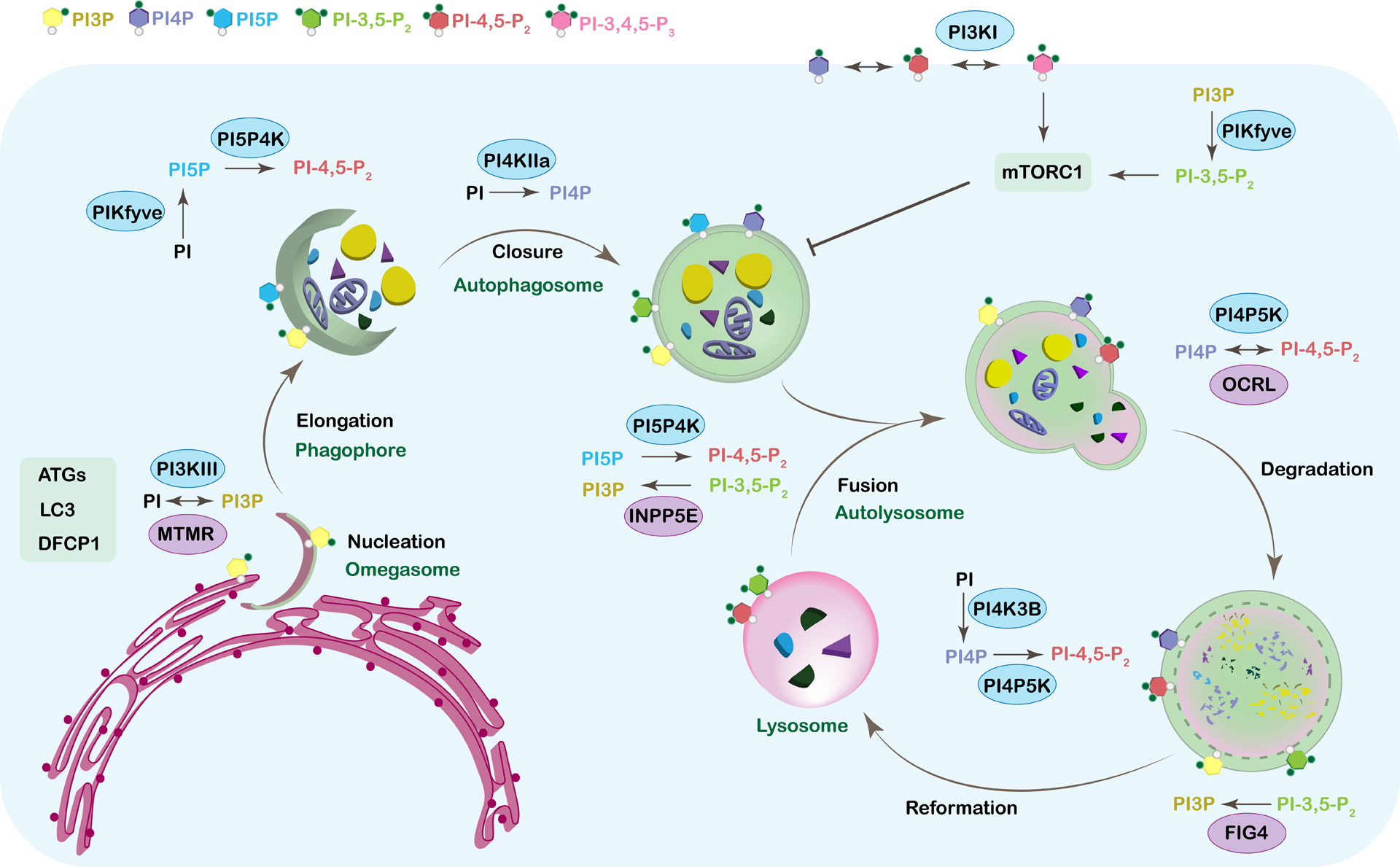
Today, the importance of autophagy in physiological processes and pathological conditions is undeniable. Initially, autophagy merely was described as an evolutionarily conserved mechanism to maintain metabolic homeostasis in times of starvation; however, in recent years it is now apparent that autophagy is a powerful regulator of many facets of cellular metabolism, that its deregulation contributes to various human pathologies, including cancer and neurodegeneration, and that its modulation has considerable potential as a therapeutic approach. Different lipid species, including sphingolipids, sterols, and phospholipids, play important roles in the various steps of autophagy. In particular, there is accumulating evidence indicating the minor group of phospholipids called the phosphoinositides as key modulators of autophagy, including the signaling processes underlying autophagy initiation, autophagosome biogenesis and maturation. In this review, we discuss the known functions to date of the phosphoinositides in autophagy and attempt to summarize the kinases and phosphatases that regulate them as well as the proteins that bind to them throughout the autophagy program. We will also provide examples of how the control of phosphoinositides and their metabolizing enzymes is relevant to understanding many human diseases.

Keywords: autophagy; lysosome; mTORC1; phosphoinositide; phosphoinositide kinase; phosphoinositide phosphatase.


Conflict of interest statement

Conflicts of Interest

The authors declare no conflict of interest.

Figures

Figure 1.
Figure 1.. Phosphoinositide cycle.
Phosphoinositides are a small group of phospholipids generated by the phosphorylation of the third, fourth and fifth positions of the inositol headgroup of phosphatidylinositol. The seven resulting phosphoinositides are cycled and regulated by a complex network of kinases and phosphatases that add or remove phosphate groups at specific positions. The precision of these enzymes is what keeps the phosphoinositide pools within a cell in perfect balance. The kinases (left) and phosphatases (right) are numbered, and the indicated number corresponds to the enzymes that allow the cycling between each of the phosphoinositide species starting with phosphatidylinositol (PI) at the center. Dashed grey arrows indicate enzymes that have not yet been characterized in vivo (Unknown).
Figure 2.
Figure 2.. Phosphoinositides in autophagy.
Autophagy is a highly regulated multistep process that is essential for recycling cytosolic components. Too much or too little autophagy leads to severe human diseases from neurodegeneration to cancer. Phosphoinositides play key roles in every step of this process from nucleation and the biogenesis of the phagophore to the reformation of the lysosome. Distinct pools of phospholipids characterize each membrane of the autophagic organelles. This phospholipid identity is ensured by specific kinases and phosphatases and their binding partners. The figure shows all the individual steps of autophagy starting with the nucleation and ending with the reformation of the lysosome. The corresponding phosphoinositide species that are known to play a role in regulating each step are shown on the membrane of the structure they are associated with and are color-coded. The kinases (blue oval) and phosphatases (purple oval) that are associated with regulating the phosphoinositide species at each step are also included. For simplicity, this model does not show all known binding partners, but these are discussed within the text.

References

    1. Balla T (2013) Phosphoinositides: tiny lipids with giant impact on cell regulation, Physiol Rev. 93, 1019–137. - PMC - PubMed
    1. Dall’Armi C, Devereaux KA & Di Paolo G (2013) The role of lipids in the control of autophagy, Curr Biol. 23, R33–45. - PMC - PubMed
    1. Klionsky DJ (2005) The molecular machinery of autophagy: unanswered questions, J Cell Sci. 118, 7–18. - PMC - PubMed
    1. Yorimitsu T & Klionsky DJ (2005) Autophagy: molecular machinery for self-eating, Cell Death Differ. 12 Suppl 2, 1542–52. - PMC - PubMed
    1. Kunz JB, Schwarz H & Mayer A (2004) Determination of four sequential stages during microautophagy in vitro, J Biol Chem. 279, 9987–96. - PubMed

Publication types

MeSH terms

Substances