Review
. 2019 Nov 5;11(11):1731.
doi: 10.3390/cancers11111731.

Carcinogenesis and Metastasis in Liver: Cell Physiological Basis

Affiliations
Review

Carcinogenesis and Metastasis in Liver: Cell Physiological Basis

Anna Rossetto et al. Cancers (Basel). .

Abstract

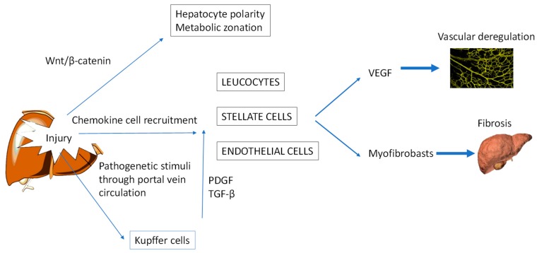
Hepatocellular carcinoma (HCC) incidence is rising. This paper summarises the current state of knowledge and recent discoveries in the cellular and physiological mechanisms leading to the development of liver cancer, especially HCC, and liver metastases. After reviewing normal hepatic cytoarchitecture and immunological characteristics, the paper addresses the pathophysiological factors that cause liver damage and predispose to neoplasia. Particular attention is given to chronic liver diseases, metabolic syndrome and the impact of altered gut microbiota, disrupted circadian rhythm and psychological stress. Improved knowledge of the multifactorial aetiology of HCC has important implications for the prevention and treatment of this cancer and of liver metastases in general.

Keywords: VEGF; circadian homeostasis; cortisol; hepatic stellate cells; hepatocellular carcinoma; immunity; myofibroblasts; nonalcoholic fatty liver disease; nonalcoholic steatohepatitis.


Conflict of interest statement

The authors declare no conflicts of interest.

Figures

Figure 1
Figure 1
Structure of hepatic lobules. Oxygenated blood enters the lobules via a hepatic arteriole, and then flows through the sinusoids and drains into a central vein in the centrilobular region. Blood enriched in nutrients and antigens from the gastrointestinal system enters the lobules via the portal vein. This blood is filtered by Kupffer cells, which are phagocytic cells found within and below the fenestrated sinusoidal endothelium. Hepatic stellate cells (fat-storing cells that secrete matrix proteins) and antigen-presenting dendritic cells are present in the space of Disse, which separates hepatocytes (epithelial liver cells) from endothelial cells. Biliary canaliculi are located between the two cords of hepatocytes.
Figure 2
Figure 2
Crosstalk among mediators of liver injury. In the absence of Wnt ligands, the Wnt/β-catenin signalling pathway is mostly inactive. During cell regenerative processes and in certain pathological conditions, the Wnt/β-catenin signal is activated. Wnt/β-catenin signalling controls several processes such as cell growth, differentiation and polarisation. Chemokines released by Kupffer cells during liver injury recruit several cell types such as leukocytes, hepatic stellate cells (HSCs) and endothelial cells to the site of injury. These cells regulate the extracellular VEGF-mediated processing of vascular permeability and angiogenesis and, through the differentiation of HSCs to myofibroblasts, promote fibrosis. Kupffer cells become activated when they phagocytise blood-borne pathogens transported by the portal circulation. Current evidence suggests that these cells are the principal effectors of fibrogenic PDGF- and TGF-β-mediated signalling, which activate HSCs and regulate extracellular matrix accumulation. VEGF, vascular endothelial growth factor; PDGF, platelet-derived growth factor; TGF-β, transforming growth factor beta.
Figure 3
Figure 3
Schematic of the liver harbouring various immune cell populations with different functions. Kupffer cells near or inside the sinusoids decrease immune reactions by releasing cytokines; if these cytokines promote inflammation, the Kupffer cells are acting as M1 macrophages; if the cytokines decrease inflammation and promote tissue repair, the cells are M2 macrophages. Dendritic cells, the main antigen-presenting cells of the immune system, are mostly present in the subcapsular space. The capsule is in contact with the viscera and peritoneum, and thus is a barrier for pathogens and neoplastic cells arriving from these sites. Neutrophils, the most abundant cell type in the blood, accumulate at sites of injury and release matrix metalloproteinases (MMPs) and reactive oxygen species (ROS), exacerbating tissue damage. Lymphocytes in the blood and bile are involved in innate and adaptive immunity and are related in the formation of ectopic germinal centre-like structures in the liver during certain pathological situations (e.g., hepatitis C virus infection). NK (natural killer) cells, natural killer cells; γδ T cells, gamma delta T cells that have a distinctive T cell receptor (TCR) composed of γδ.
Figure 4
Figure 4
The gut–liver axis. The gut microbiota exacerbates liver diseases by: (i) the production of prostaglandins which suppress tumour immunity; (ii) the translocation of bacterial lipopolysaccharides (LPS) and other metabolites, particularly indoles, to the liver and other organs during states of high intestinal permeability; and (iii) the alterations of bile acid composition, leading to the accumulation of secondary bile acids in the liver.
Figure 5
Figure 5
Possible causes of liver damage due to circadian rhythm alterations, hypoxia and psychological stress. Disruption of the circadian rhythm is a risk factor for obesity, metabolic disorders and HCC. Hypoxia-inducible factors (HIFs) control gene expression during hypoxia, enabling cells to survive in a hypoxic environment. HIFs reprogramme the cell’s metabolism towards anaerobic glycolysis to increase energy through ATP production, thus lowering the pH value of tissue. Low pH activates the inflammatory program of monocytes, reduces the number and function of T cells, and promotes angiogenesis. Chronic psychological stress leads to a cortisol secretory burst, which creates an immunosuppressive state in the liver.

References

    1. Ghouri Y.A., Mian I., Rowe J.H. Review of hepatocellular carcinoma: Epidemiology, etiology, and carcinogenesis. J. Carcinog. 2017;16:1. - PMC - PubMed
    1. Chang M.H., Chen C.J., Lai M.S., Hsu H.M., Wu T.C., Kong M.S., Liang D.C., Shau W.Y., Chen D.S. Universal hepatitis B vaccination in Taiwan and the incidence of hepatocellular carcinoma in children. Taiwan Childhood Hepatoma Study Group. N. Engl. J. Med. 1997;336:1855–1859. doi: 10.1056/NEJM199706263362602. - DOI - PubMed
    1. Carrat F., Fontaine H., Dorival C., Simony M., Diallo A., Hezode C., De Ledinghen V., Larrey D., Haour G., Bronowicki J.-P., et al. Clinical outcomes in patients with chronic hepatitis C after direct-acting antiviral treatment: A prospective cohort study. Lancet. 2019;393:1453–1464. doi: 10.1016/S0140-6736(18)32111-1. - DOI - PubMed
    1. Meringer H., Shibolet O., Deutsch L. Hepatocellular carcinoma in the post-hepatitis C virus era: Should we change the paradigm? World J. Gastroenterol. 2019;25:3929–3940. doi: 10.3748/wjg.v25.i29.3929. - DOI - PMC - PubMed
    1. Schütte K., Schulz C., Poranzke J., Antweiler K., Bornschein J., Bretschneider T., Arend J., Ricke J., Malfertheiner P. Characterization and prognosis of patients with hepatocellular carcinoma (HCC) in the non-cirrhotic liver. BMC Gastroenterol. 2014;14:117. doi: 10.1186/1471-230X-14-117. - DOI - PMC - PubMed

LinkOut - more resources