

Spontaneous regression of extradural intraspinal cysts in a dog: a case report
- PMID: 31694633
- PMCID: PMC6833175
- DOI: 10.1186/s12917-019-2152-x
Spontaneous regression of extradural intraspinal cysts in a dog: a case report
Abstract
Background: Extradural intraspinal cysts are fluid accumulations that appear to be associated with increased motion at vertebral joints.
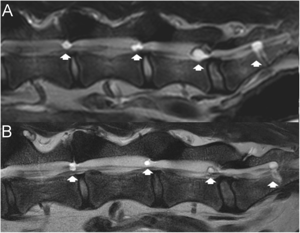
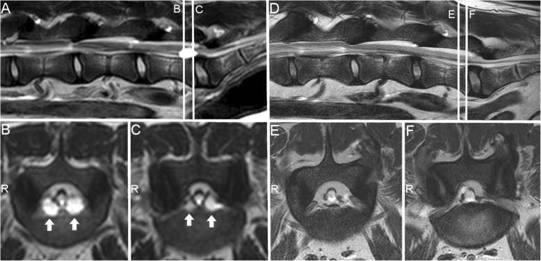
Case presentation: We report the spontaneous regression of lumbar and lumbosacral cysts (presumably synovial cysts) and the unusual occurrence of an S1-2 extradural intraspinal cyst in a dog. The dog presented with lumbosacral pain. Six extradural intraspinal cysts were observed on high-field magnetic resonance imaging from L5-6 to S1-S2. The cysts between L5-6 and L7-S1 ranged from 0.12 to 0.44cm2 at their largest area. The largest cyst was located at S1-2 (left), measuring 0.84 cm2 at its largest view. The dog was medically managed. A follow-up magnetic resonance imaging scan was obtained 3.5 years after the first imaging. All cysts except the one at S1-2 had reduced in size. Mean reduction in size was 59.6% (35-81%).
Conclusions: In summary, we report a case with multiple extradural intraspinal cysts that underwent spontaneous regression of all but one cyst during a 3.5-year follow-up period. Whether this is a single occurrence, or is part of the natural history of these cysts in the lumbosacral region of dogs, remains to be established. Spontaneous regression of intraspinal cysts had not been described in dogs.
Keywords: Juxtafacet cysts; Lumbar; Magnetic resonance imaging; Synovial cyst.
Conflict of interest statement
The authors declare that they have no competing interests.
Figures
References
-
- Lowrie ML, Platt SR, Garosi LS. Extramedullary spinal cysts in dogs. Vet Surg. 2014;43:650–662. - PubMed
Publication types
MeSH terms
LinkOut - more resources
Full Text Sources
