. 2019 Dec 9;6(6):ENEURO.0429-19.2019.
doi: 10.1523/ENEURO.0429-19.2019. Print 2019 Nov/Dec.

Threat Anticipation in Pulvinar and in Superficial Layers of Primary Visual Cortex (V1). Evidence from Layer-Specific Ultra-High Field 7T fMRI

Affiliations

Threat Anticipation in Pulvinar and in Superficial Layers of Primary Visual Cortex (V1). Evidence from Layer-Specific Ultra-High Field 7T fMRI

Ai Koizumi et al. eNeuro. .

Abstract

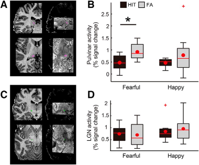
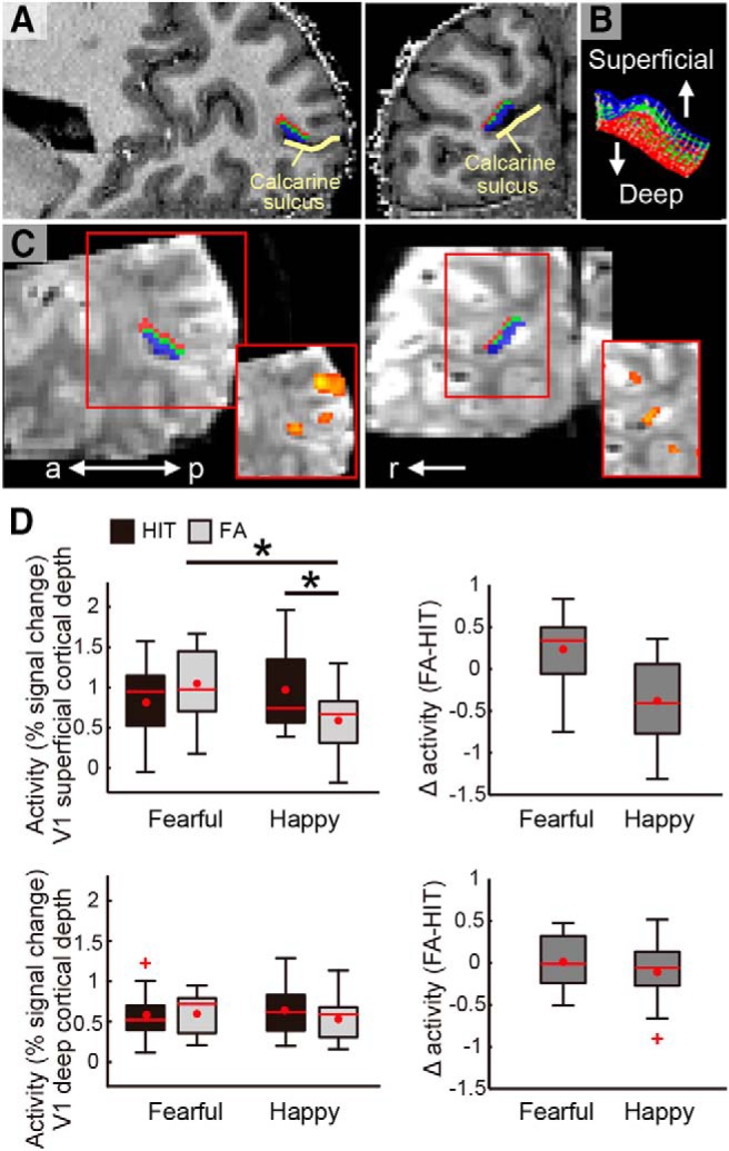
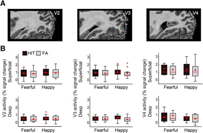
The perceptual system gives priority to threat-relevant signals with survival value. In addition to the processing initiated by sensory inputs of threat signals, prioritization of threat signals may also include processes related to threat anticipation. These neural mechanisms remain largely unknown. Using ultra-high-field 7 tesla (7T) fMRI, we show that anticipatory processing takes place in the early stages of visual processing, specifically in the pulvinar and V1. When anticipation of a threat-relevant fearful face target triggered false perception of not-presented target, there was enhanced activity in the pulvinar as well as in the V1 superficial-cortical-depth (layers 1-3). The anticipatory activity was absent in the LGN or higher visual cortical areas (V2-V4). The effect in V1 was specific to the perception of fearful face targets and did not generalize to happy face targets. A preliminary analysis showed that the connectivity between the pulvinar and V1 superficial-cortical-depth was enhanced during false perception of threat, indicating that the pulvinar and V1 may interact in preparation of anticipated threat. The anticipatory processing supported by the pulvinar and V1 may play an important role in non-sensory-input-driven anxiety states.

Keywords: 7 tesla fMRI; V1 cortical layer; fearful face; pulvinar; threat perception.


Figures

Figure 1.
Figure 1.
Design of the fearful face detection task. In each trial, either a fearful face target or a neutral face was presented briefly, followed by a mask consisting of a neutral face with a different identity than the target face. Participants responded whether they perceived a fearful target or neutral face by pressing the response key, which was randomly assigned trial-wise. The control task used happy face targets and neutral faces (images not shown), and otherwise identical procedures. ITI: intertrial interval. See also Extended Data Figure 1-1.
Figure 2.
Figure 2.
Activity in the pulvinar and LGN. A, Demonstrations of pulvinar ROI from a representative participant, shown on the anatomic image (left panels) and EPI (right panels). B, Pulvinar showed enhanced activity in FA trials relative to HIT trials during the fearful face detection task (t(10) = –2.94, p = 0.015) but not during the happy face detection task (t(10) = –1.23, p = 0.247). There was no significant interaction between percept type and emotion (F(1,10) = 0.16, p = 0.702). C, Demonstrations of the LGN ROI from an example participant. D, Unlike the pulvinar, the LGN showed no differential activity between the percept types and facial emotions. Box plot shows upper (75%) and lower (25%) quartiles with median (red line) and mean (red dot), with whisker showing maximum and minimum value. An outlier (outside of ±2.7 SDs within a distribution for a given condition) is shown with a red cross; *p < 0.05. See also Extended Data Figures 2-1A, 2-2.
Figure 3.
Figure 3.
Demonstrations of V1 cortical depths and cortical depth dependent activity during the tasks. A, V1 cortical depth is visualized on an anatomic image of a representative participant with sagittal (left panel) and coronal views (right panel). The voxels allocated to superficial (outwards to pial surface) and deep (inwards to white matter) cortical depths are shown in blue and red, respectively. The voxels allocated to the intermediate depth (shown in green) were disregarded in the main analyses (see Materials and Methods). B, The cortical grid mesh within which the voxels were allocated. C, The V1 cortical depth is visualized on EPI images (visualized in 3D for demonstrative purpose) in sagittal and coronal views (left and right, respectively). Red squares at the lower right demonstrate activity for all face targets relative to baseline on the EPI smoothed with a 3D kernel of 2.4-mm FWHM. D, Peak activity at V1 superficial and deep cortical depths (upper and lower rows, respectively) in HIT and FA trials in the fearful face and happy face detection tasks. The difference in activity between FA and HIT trials are demonstrated in the right panel for each task, with a larger value indicating greater activity for FA than for HIT trials. Box plot shows upper (75%) and lower (25%) quartiles with median (red line) and mean (red dot), with whisker showing maximum and minimum value. An outlier (outside of ±2.7 SDs within a distribution for a given condition) is shown with a red cross. a: anterior, p: posterior, r: right; *p < 0.05. See also Extended Data Figures 2-1B, 3-1, 3-2, 3-3, 3-4.
Figure 4.
Figure 4.
Control analyses showing no differential activity in V2, V3, and V4 in HIT compared with FA trials. A, V2, V3, and V4 cortical depths are visualized on the anatomic image of an example participant, with the same color coding as for V1 shown in Figure 3. B, Unlike V1, V2–V4 did not show any differential activity between the percept types and facial emotions, regardless of cortical depth. Box plot shows upper (75%) and lower (25%) quartiles with median (red line) and mean (red dot), with whisker showing maximum and minimum value. An outlier (outside of ±2.7 SDs within a distribution for a given condition) is shown with a red cross.

Similar articles

Cited by

References

    1. Andersson JL, Skare S, Ashburner J (2003) How to correct susceptibility distortions in spin-echo echo-planar images: application to diffusion tensor imaging. Neuroimage 20:870–888. 10.1016/S1053-8119(03)00336-7 - DOI - PubMed
    1. Bishop SJ (2007) Neurocognitive mechanisms of anxiety: an integrative account. Trends Cogn Sci 11:307–316. 10.1016/j.tics.2007.05.008 - DOI - PubMed
    1. Boynton GM, Engel SA, Glover GH, Heeger DJ (1996) Linear systems analysis of functional magnetic resonance imaging in human V1. J Neurosci 16:4207–4221. - PMC - PubMed
    1. Brainard DH (1997) The psychophysics toolbox. Spat Vis 10:433–436. - PubMed
    1. Bridge H, Leopold DA, Bourne JA (2016) Adaptive pulvinar circuitry supports visual cognition. Trends Cogn Sci 20:146–157. 10.1016/j.tics.2015.10.003 - DOI - PMC - PubMed

Publication types

LinkOut - more resources