. 2019 Dec 6;14(12):1719-1732.
doi: 10.2215/CJN.05830519. Epub 2019 Nov 6.

Complement Activation and Thrombotic Microangiopathies

Affiliations

Complement Activation and Thrombotic Microangiopathies

Marta Palomo et al. Clin J Am Soc Nephrol. .

Abstract

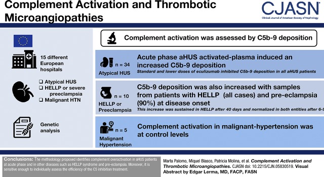
Background and objectives: Atypical hemolytic uremic syndrome is a form of thrombotic microangiopathy caused by dysregulation of the alternative complement pathway. There is evidence showing complement activation in other thrombotic microangiopathies. The aim of this study was to evaluate complement activation in different thrombotic microangiopathies and to monitor treatment response.

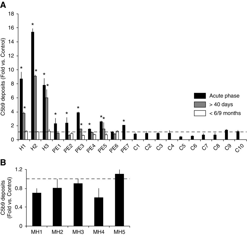
Design, setting, participants, & measurements: Complement activation was assessed by exposing endothelial cells to sera or activated-patient plasma-citrated plasma mixed with a control sera pool (1:1)-to analyze C5b-9 deposits by immunofluorescence. Patients with atypical hemolytic uremic syndrome (n=34) at different stages of the disease, HELLP syndrome (a pregnancy complication characterized by hemolysis, elevated liver enzymes, and low platelet count) or severe preeclampsia (n=10), and malignant hypertension (n=5) were included.

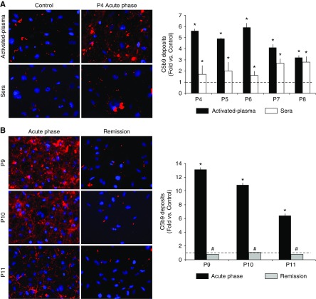
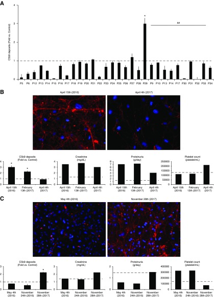
Results: Acute phase atypical hemolytic uremic syndrome-activated plasma induced an increased C5b-9 deposition on endothelial cells. Standard and lower doses of eculizumab inhibited C5b-9 deposition in all patients with atypical hemolytic uremic syndrome, except in two who showed partial remission and clinical relapse. Significant fibrin formation was observed together with C5b-9 deposition. Results obtained using activated-plasma samples were more marked and reproducible than those obtained with sera. C5b-9 deposition was also increased with samples from patients with HELLP (all cases) and preeclampsia (90%) at disease onset. This increase was sustained in those with HELLP after 40 days, and levels normalized in patients with both HELLP and preeclampsia after 6-9 months. Complement activation in those with malignant hypertension was at control levels.

Conclusions: The proposed methodology identifies complement overactivation in patients with atypical hemolytic uremic syndrome at acute phase and in other diseases such as HELLP syndrome and preeclampsia. Moreover, it is sensitive enough to individually assess the efficiency of the C5 inhibition treatment.

Keywords: HELLP syndrome; alternative; antibodies; atypical hemolytic uremic syndrome; complement C9; complement activation; complement membrane; complement pathway; complement system proteins; eculizumab; endothelial cells; female; fibrin; fluorescent antibody technique; humanized; humans; hypertension; malignant; monoclonal; pre-eclampsia; pregnancy; recurrence; thrombotic microangiopathies.


Figures

None
Graphical abstract
Figure 1.
Figure 1.
Atypical hemolytic uremic syndrome (aHUS)–activated plasma induces C5b-9 deposition on microvascular endothelial cells with a coefficient of variation significantly lower than that obtained using aHUS serum. (A) Representative microscopy image of C5b-9 deposition staining (red) on endothelial cells (4′,6-diamidino-2-phenylindole–stained nuclei, blue) induced by activated plasma or sera from a control individual and a patient with aHUS in the acute phase (patient 4, P4). Bar diagram represents quantification of C5b-9 deposits expressed as fold increase of the covered surface with respect to the control, from five different patients with aHUS in the acute phase (P4 to P8). Black bars represent the results obtained with activated plasma and white bars with serum. The dotted line represents control values. (B) Representative microscopy images of staining of C5b-9 deposition on endothelial cells (induced by activated plasma from the acute phase and at remission in three patients: P9 to P11). Bar diagram represents C5b-9 quantification, black bars represent results from the acute phase, and gray bars represent remission. All values at the acute phase were statistically higher than in control samples (*P<0.05) and all values at remission are statistically lower than those at the acute phase (#P<0.05). Statistical analysis was performed with raw data using the t test for paired samples.
Figure 2.
Figure 2.
C5b-9 deposits on activated-endothelial cells are at control level or even lower when analyzing the effect of activated-plasma samples from aHUS patients treated at regular doses of eculizumab or lower doses. Identification of two patients with partial remision and clinical relapse. (A) Bar diagrams show the results of C5b-9 deposition on endothelial cells of activated plasma from 20 patients with aHUS (P5 to P29) who were treated at regular doses of eculizumab (according to weight in pediatric patients; 900 mg/wk for 4 weeks as induction therapy for adults, and 1200 mg every 2 weeks thereafter as maintenance). All values resulted in control levels except in patient 29, in whom they were significantly higher (*). Bar diagram also shows, under the continuous line and ## symbol, the results from ten patients with aHUS who were treated at lower doses or for longer intervals than those specified in the data sheet. Activated plasma from those patients induced C5b-9 deposition at similar or even lower levels than control activated plasma. The dotted line represents control values. Statistical analysis was performed with raw data using the t test for paired samples. (B) Results from the C5b-9 deposition assay allowed the identification of a patient who was receiving an underdose of eculizumab and allowed for a consequent dose adjustment. The left image corresponds to a representative image of C5b-9 deposits induced by activated plasma from patient 29 which was obtained on April 15, 2016. At that time point, C5b-9 deposition was statistically higher (*P<0.05) than control levels as seen in the bar diagrams. The right image corresponds to activated plasma from the same patient obtained on April 4, 2016, after several eculizumab dose changes, showing a negative result. Statistical analysis was performed with raw data using the t test for paired samples. The results from C5b-9 assays at three different time points corresponding to different eculizumab guidelines are quantified in the bar diagrams along with creatinine (mg/dl), proteinuria (g/d), and platelet count (platelet/ml). (C) Results from the C5b-9 deposition assay confirmed the identification of a patient with aHUS recurrence (left image) when C5b-9 deposition was higher than control levels (bar diagrams), and complete clinical recovery (right image) after the start of eculizumab treatment. Bar diagrams show C5b-9 quantification, creatinine (mg/dl), proteinuria (g/d), and platelet count (platelet/ml) at different time points.
Figure 3.
Figure 3.
Activated plasma from patients with HELLP syndrome and preeclampsia at onset induced an increase C5b-9 deposition on endothelial cells. (A) Quantification of C5b-9 deposits on endothelial cells show that three HELLP syndrome patients (H1 to H3) showed marked complement activation in the acute phase (black bars) that was still increased after 40 days (gray bars), and normalized after 6–9 months (white bars). All except one patient with preeclampsia (PE) also showed increased complement activation in the acute phase, but only two were still positive after 40 days. All patients with preeclampsia analyzed had normal complement activity after 6–9 months. Ten healthy pregnant women were also analyzed (C1 to C10), obtaining normal C5b-9 deposits. The dotted line represents control values. *P<0.05 represents values statistically higher than control samples. Statistical analysis was performed with raw data using the t test for paired samples. (B) Bar diagrams show results from C5b-9 deposition induced by activated plasma from five patients with malignant hypertension (MH1 to MH5). All patients in the acute phase showed normal complement activity, represented by the dotted line. Statistical analysis was performed with raw data using the t test for paired samples.

Comment in

References

    1. George JN, Nester CM: Syndromes of thrombotic microangiopathy. N Engl J Med 371: 654–666, 2014 - PubMed
    1. Sethi S, Fervenza FC: Pathology of renal diseases associated with dysfunction of the alternative pathway of complement: C3 glomerulopathy and atypical hemolytic uremic syndrome (aHUS). Semin Thromb Hemost 40: 416–421, 2014 - PubMed
    1. Román E, Mendizábal S, Jarque I, de la Rubia J, Sempere A, Morales E, Praga M, Ávila A, Górriz JL: Secondary thrombotic microangiopathy and eculizumab: A reasonable therapeutic option. Nefrologia 37: 478–491, 2017 - PubMed
    1. Cavero T, Rabasco C, López A, Román E, Ávila A, Sevillano Á, Huerta A, Rojas-Rivera J, Fuentes C, Blasco M, Jarque A, García A, Mendizabal S, Gavela E, Macía M, Quintana LF, María Romera A, Borrego J, Arjona E, Espinosa M, Portolés J, Gracia-Iguacel C, González-Parra E, Aljama P, Morales E, Cao M, Rodríguez de Córdoba S, Praga M: Eculizumab in secondary atypical haemolytic uraemic syndrome. Nephrol Dial Transplant 32: 466–474, 2017 - PMC - PubMed
    1. Jokiranta TS: HUS and atypical HUS. Blood 129: 2847–2856, 2017 - PMC - PubMed

Publication types

MeSH terms

Substances