. 2020 Jan 17;94(3):e01034-19.
doi: 10.1128/JVI.01034-19. Print 2020 Jan 17.

Genetic Diversity of Collaborative Cross Mice Controls Viral Replication, Clinical Severity, and Brain Pathology Induced by Zika Virus Infection, Independently of Oas1b

Affiliations

Genetic Diversity of Collaborative Cross Mice Controls Viral Replication, Clinical Severity, and Brain Pathology Induced by Zika Virus Infection, Independently of Oas1b

Caroline Manet et al. J Virol. .

Abstract

The explosive spread of Zika virus (ZIKV) has been associated with major variations in severe disease and congenital afflictions among infected populations, suggesting an influence of host genes. We investigated how genome-wide variants could impact susceptibility to ZIKV infection in mice. We first describe that the susceptibility of Ifnar1-knockout mice is largely influenced by their genetic background. We then show that Collaborative Cross (CC) mice, which exhibit a broad genetic diversity, in which the type I interferon receptor (IFNAR) was blocked by an anti-IFNAR antibody expressed phenotypes ranging from complete resistance to severe symptoms and death, with large variations in the peak and the rate of decrease in the plasma viral load, in the brain viral load, in brain histopathology, and in the viral replication rate in infected cells. The differences in susceptibility to ZIKV between CC strains correlated with the differences in susceptibility to dengue and West Nile viruses between the strains. We identified highly susceptible and resistant mouse strains as new models to investigate the mechanisms of human ZIKV disease and other flavivirus infections. Genetic analyses revealed that phenotypic variations are driven by multiple genes with small effects, reflecting the complexity of ZIKV disease susceptibility in the human population. Notably, our results rule out the possibility of a role of the Oas1b gene in the susceptibility to ZIKV. Altogether, the findings of this study emphasize the role of host genes in the pathogeny of ZIKV infection and lay the foundation for further genetic and mechanistic studies.IMPORTANCE In recent outbreaks, ZIKV has infected millions of people and induced rare but potentially severe complications, including Guillain-Barré syndrome and encephalitis in adults. While several viral sequence variants were proposed to enhance the pathogenicity of ZIKV, the influence of host genetic variants in mediating the clinical heterogeneity remains mostly unexplored. We addressed this question using a mouse panel which models the genetic diversity of the human population and a ZIKV strain from a recent clinical isolate. Through a combination of in vitro and in vivo approaches, we demonstrate that multiple host genetic variants determine viral replication in infected cells and the clinical severity, the kinetics of blood viral load, and brain pathology in mice. We describe new mouse models expressing high degrees of susceptibility or resistance to ZIKV and to other flaviviruses. These models will facilitate the identification and mechanistic characterization of host genes that influence ZIKV pathogenesis.

Keywords: Collaborative Cross; Zika; Zika virus; flavivirus; genetic diversity; host genetics; mouse model.


Figures

FIG 1
FIG 1
ZIKV disease severity in Ifnar1-deficient mice is driven by the genetic background. Six- to 7-week-old 129-Ifnar1 (n = 7) and B6-Ifnar1 (n = 10) mice were infected i.p. with 107 FFUs of ZIKV FG15 and monitored for 14 days. (A) Average clinical score, with numerical values given as follows: 0, no symptoms; 1, ruffled fur; 2, emaciation, hunched posture, and/or hypoactivity; 3, hind limb weakness, prostration, and/or closed eyes; and 4, moribund or dead. (B) Kaplan-Meier survival curves showing 100% lethality in B6-Ifnar1 mice at day 7 p.i. and the survival of 6/7 129-Ifnar1 mice, ***, P = 0.0002 (log-rank test). B6-Ifnar1 mice developed early symptoms, which rapidly evolved to death, while 129-Ifnar1 mice developed symptoms 2 days later, which eventually resolved in most mice.
FIG 2
FIG 2
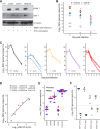
Establishment and validation of the experimental conditions for assessing susceptibility to ZIKV in CC strains. (A) The efficacy of the MAR1-5A3 MAb (100 μg for 5 × 10⁶ cells) at blocking the IFNAR receptor in diverse mouse genetic backgrounds was determined by assessing STAT1 phosphorylation (pStat 1) by Western blotting on mouse embryonic fibroblasts (MEFs) derived from the C57BL/6J, CC001, CC071, and CD-1 strains. (B) Plasma viral load, measured on days 2, 3, and 6 p.i. by RT-qPCR, in 6- to 8-week-old mice of the CC001 and CC071 strains that had been treated with MAb MAR1-5A3 24 h prior to ZIKV infection (filled circles; n = 9 and 8, respectively) or untreated (open circles; n = 3 and 2, respectively). x, a sample with a level below the detection level. (C) Kinetics of the plasma viral load in 129-Ifnar1 mice and 4 CC strains measured by RT-qPCR. Each circle represents the result for a 6- to 8-week-old mouse analyzed on days 1, 2, 3, and 6. (D) Correlation between the plasma viral load determined by FFA (x axis) and RT-qPCR (y axis) in 46 blood samples from 129-Ifnar1 and B6-Ifnar1 mice and 10 CC strains (circles show the mean for each strain; the number of 6- to 8-week-old mice per strain is shown in parentheses). (E) Plasma viral load, measured by RT-qPCR at day 2 p.i., in 6- to 8-week-old males and females of the 129-Ifnar1 strain and of 4 CC strains (n ≥ 4 mice per group). (F) Effect of the dose of MAR1-5A3 antibody treatment on the rate of decrease of the plasma viral load. Seven- to 9-week-old mice of the CC012 and CC037 strains (4 mice per group) received 2 mg of the MAR1-5A3 MAb 1 day prior to being inoculated i.p. with 107 FFUs of ZIKV FG15. The groups receiving 4 mg of the MAb received additional i.p. injections of 1 mg of MAb on days 2 and 4 p.i. The plasma viral load was measured on days 2 and 6 p.i. (and the results are presented above and below the dashed line, respectively).
FIG 3
FIG 3
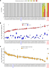
CC genetic diversity strongly impacts the clinical severity of infection and the plasma viral load. Thirty-five CC strains (n = 2 to 9 6- to 8-week-old mice per strain) were infected i.p. with 107 FFUs of ZIKV FG15 at 24 h after i.p. injection of 2 mg of the MAR1-5A3 MAb. 129-Ifnar1 (n = 24) and B6-Ifnar1 (n = 5) mice were similarly infected without MAb treatment. (A) Clinical scores at day 7 p.i. as the percentage of mice in the five levels of severity (as described in the legend to Fig. 1). (B) Plasma viral load at days 2 p.i. (upper values) and 6 p.i. (lower values) quantified by RT-qPCR, shown as box-whisker plots, with outliers shown as dots (the strains are shown in the same order in which they are shown in panel A). (C) Difference between plasma viral loads at days 2 and 6 p.i. Strains are sorted by increasing absolute difference and are therefore in an order different from that in panels A and B. (B and C) Arrows indicate the subset of CC mouse strains selected for detailed study.
FIG 4
FIG 4
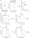
The differences in susceptibility to ZIKV between CC strains correlate with the differences in susceptibility to other flaviviruses. (A) Six- to 8-week-old mice from three selected CC strains treated with the MAR1-5A3 MAb and 129-Ifnar1 mice were infected intraperitoneally with 103 FFUs of ZIKV HD78788. (Left) Average clinical score, with numerical values given as described in the legend to Fig. 1; (center) Kaplan-Meier survival curves (log-rank test); (right) plasma viral load at day 2 p.i., measured by RT-qPCR (P values were determined by the Wilcoxon rank-sum test). (B) The viral loads after ZIKV infection (left; data extracted from Fig. 3) and DENV infection (right; data for i.v. infection with 2 × 10⁶ FFUs of DENV KDH0026A) in MAb-treated 6- to 8-week-old CC001, CC071, and B6 mice and in 129-Ifnar1 and B6-Ifnar1 were compared (P values were determined by the t test). (C) (Left) Kaplan-Meier survival curves for four 8- to 12-week-old male mice of each of the three selected CC strains infected i.p. with 1,000 FFUs of WNV strain IS-98-ST1 and monitored for 14 days (P values were determined by the log-rank test); (right) Kaplan-Meier survival curves for four to five 8- to 12-week-old male mice of the BALB/cByJ strain and each of the three selected CC strains infected i.p. with 100 PFU of RVFV strain ZH548 and monitored for 14 days (log-rank test, P > 0.05). *, P < 0.05; **, P < 0.01; ***, P < 0.001.
FIG 5
FIG 5
Genetic analysis of susceptibility to ZIKV fails to identify simple genetic control. Genome-wide linkage analysis for the plasma viral load at day 2 p.i. (A), the plasma viral load at day 6 p.i. (B), and the rate of decrease in the plasma viral load (C) for the 35 CC strains for which the results are shown in Fig. 3. The x axis represents the genomic location; the y axis is the LOD score, representing the statistical association between the phenotype and the genomic location. Genome-wide thresholds of P equal to 0.1, 0.05, and 0.01, computed from 1,000 permutations, are represented by dashed black, dashed red, and plain red lines, respectively. No genome location reached the threshold of P equal to 0.05.
FIG 6
FIG 6
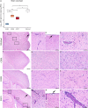
Genetic variations between CC strains control the brain viral load and histological profile in infected mice. Four to five 6- to 8-week-old mice of the 129-Ifnar1 strain and three selected CC strains were infected i.p. with 107 FFUs of ZIKV FG15 24 h after i.p. injection of 2 mg of the MAR1-5A3 MAb. (A) Brain viral load measured by RT-qPCR at day 6 p.i. *, P < 0.05, Wilcoxon rank-sum test. (B to N) Representative HE-stained brain sections at three different magnifications. (B to E) 129-Ifnar1 mice (n = 3). Black rectangle, encephalitis with perivascular lymphocyte cuffs. (B and D) Arrows, lesions of subacute leptomeningoencephalitis. (C) Arrows, perivascular lymphocyte cuffs. (F to H) CC001 mice (n = 5). (I to K) CC005 mice (n = 5). Black rectangle, encephalitis with perivascular lymphocyte cuffs; arrow, perivascular cuffing. (L to N) CC071 mice (n = 4).
FIG 7
FIG 7
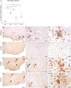
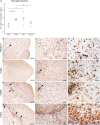
Genetic variations between CC strains control brain neuroinflammation in infected mice. Microglial reactivity was assessed on brain sections from the same mice described in the legend to Fig. 6 by anti-Iba1 immunohistochemistry. (A) Quantification of the Iba1 labeling signal on the brain sections. P values were determined by t tests. *, P < 0.05; **, P < 0.01; ***, P < 0.001. (B to M) Representative anti-Iba1 immunohistochemistry of brain sections at three different magnifications. (B to D) 129-Ifnar1 mice (n = 3); (E to G) CC001 mice (n = 5); (H to J) CC005 mice (n = 5); (K to M) CC071 mice (n = 4). Arrowheads, nodules of activated microglial cells.
FIG 8
FIG 8
Intracranial ZIKV FG15 infection results in a strain-dependent viral load and brain histological lesions. Groups of 5- to 6-week-old mice of the 129-Ifnar1 strain and three selected CC strains (3 to 5 mice per strain) were infected i.c. with 105 FFUs of ZIKV FG15 in the absence of prior anti-IFNAR treatment. (A) Brain viral load measured by RT-qPCR at day 6 p.i. P values were determined by the Wilcoxon rank-sum test. *, P < 0.05; **, P < 0.01. (B to O) Representative HE-stained brain sections at three different magnifications. (B to E) 129-Ifnar1 mice (n = 4). Black rectangles, encephalitis with perivascular lymphocyte cuffs; arrows, leptomeningitis. (F to H) CC001 mice (n = 5). (I to K) CC005 mice (n = 4). (L to O) CC071 mice (n = 5). Black rectangles, encephalitis with perivascular lymphocyte cuffs; arrows, leptomeningitis.
FIG 9
FIG 9
Intracranial ZIKV FG15 infection results in strain-dependent neuroinflammation of the brain. Microglial reactivity was assessed on brain sections from the same mice described in the legend to Fig. 8 by anti-Iba1 immunohistochemistry. (A) Quantification of the Iba1 labeling signal on the brain sections. P values were determined by t tests. *, P < 0.05; **, P < 0.01; ***, P < 0.001. (B to M) Representative anti-Iba1 immunohistochemistry of brain sections at three different magnifications. (B to D) 129-Ifnar1 mice (n = 4); (E to G) CC001 mice (n = 5); (H to J) CC005 mice (n = 4); (K to M) CC071 mice (n = 5). Arrowheads, nodules of activated microglial cells.
FIG 10
FIG 10
Enhanced ZIKV replication in CC071 MEFs compared with CC001 MEFs. MEFs derived from CC001 and CC071 mouse embryos were infected with ZIKV FG15 at an MOI of 5. The ZIKV titer in the supernatant was quantified by a focus-forming assay at 24, 48, and 72 h p.i. The data represent the mean ± SEM from 3 biological replicates. The results were replicated in 3 independent experiments (t tests, ***, P < 0.001).

References

    1. Petersen LR, Jamieson DJ, Powers AM, Honein MA. 2016. Zika virus. N Engl J Med 374:1552–1563. doi:10.1056/NEJMra1602113. - DOI - PubMed
    1. Talero-Gutierrez C, Rivera-Molina A, Perez-Pavajeau C, Ossa-Ospina I, Santos-Garcia C, Rojas-Anaya MC, de-la-Torre A. 2018. Zika virus epidemiology: from Uganda to world pandemic, an update. Epidemiol Infect 146:673–679. doi:10.1017/s0950268818000419. - DOI - PMC - PubMed
    1. Cao-Lormeau V-M, Blake A, Mons S, Lastère S, Roche C, Vanhomwegen J, Dub T, Baudouin L, Teissier A, Larre P, Vial A-L, Decam C, Choumet V, Halstead SK, Willison HJ, Musset L, Manuguerra J-C, Despres P, Fournier E, Mallet H-P, Musso D, Fontanet A, Neil J, Ghawché F. 2016. Guillain-Barre syndrome outbreak associated with Zika virus infection in French Polynesia: a case-control study. Lancet 387:1531–1539. doi:10.1016/S0140-6736(16)00562-6. - DOI - PMC - PubMed
    1. Munoz LS, Parra B, Pardo CA. 2017. Neurological implications of Zika virus infection in adults. J Infect Dis 216:S897–S905. doi:10.1093/infdis/jix511. - DOI - PMC - PubMed
    1. Rasmussen SA, Jamieson DJ, Honein MA, Petersen LR. 2016. Zika virus and birth defects—reviewing the evidence for causality. N Engl J Med 374:1981–1987. doi:10.1056/NEJMsr1604338. - DOI - PubMed

Publication types

MeSH terms

Substances