. 2020 Jan 6;94(2):e01439-19.
doi: 10.1128/JVI.01439-19. Print 2020 Jan 6.

Near-Atomic-Resolution Cryo-Electron Microscopy Structures of Cucumber Leaf Spot Virus and Red Clover Necrotic Mosaic Virus: Evolutionary Divergence at the Icosahedral Three-Fold Axes

Affiliations

Near-Atomic-Resolution Cryo-Electron Microscopy Structures of Cucumber Leaf Spot Virus and Red Clover Necrotic Mosaic Virus: Evolutionary Divergence at the Icosahedral Three-Fold Axes

Michael B Sherman et al. J Virol. .

Abstract

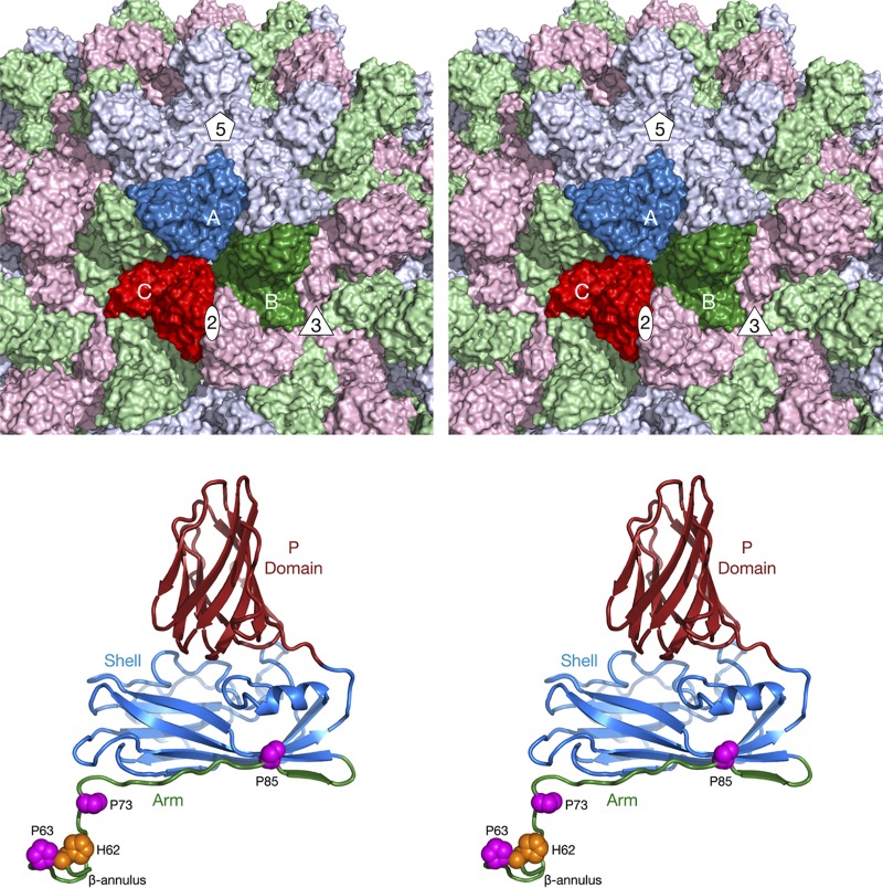
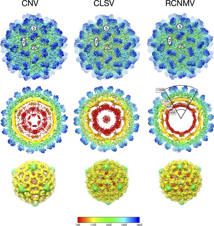
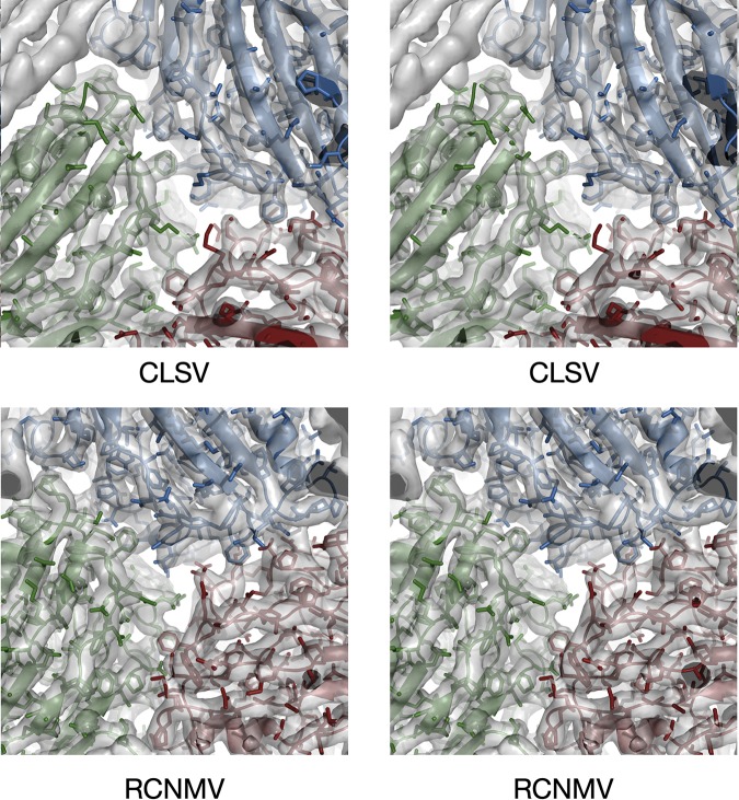
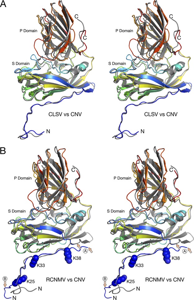
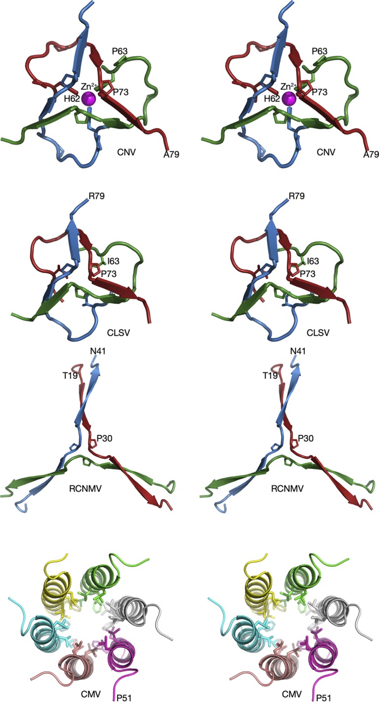
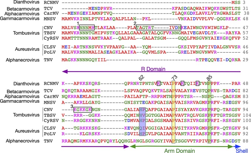
Members of the Tombusviridae family have highly similar structures, and yet there are important differences among them in host, transmission, and capsid stabilities. Viruses in the Tombusviridae family have single-stranded RNA (ssRNA) genomes with T=3 icosahedral protein shells with a maximum diameter of ∼340 Å. Each capsid protein is comprised of three domains: R (RNA binding), S (shell), and P (protruding). Between the R domain and S domain is the "arm" region that studies have shown to play a critical role in assembly. To better understand how the details of structural differences and similarities influence the Tombusviridae viral life cycles, the structures of cucumber leaf spot virus (CLSV; genus Aureusvirus) and red clover necrotic mosaic virus (RCNMV; genus Dianthovirus) were determined to resolutions of 3.2 Å and 2.9 Å, respectively, with cryo-electron microscopy and image reconstruction methods. While the shell domains had homologous structures, the stabilizing interactions at the icosahedral 3-fold axes and the R domains differed greatly. The heterogeneity in the R domains among the members of the Tombusviridae family is likely correlated with differences in the sizes and characteristics of the corresponding genomes. We propose that the changes in the R domain/RNA interactions evolved different arm domain interactions at the β-annuli. For example, RCNMV has the largest genome and it appears to have created the necessary space in the capsid by evolving the shortest R domain. The resulting loss in RNA/R domain interactions may have been compensated for by increased intersubunit β-strand interactions at the icosahedral 3-fold axes. Therefore, the R and arm domains may have coevolved to package different genomes within the conserved and rigid shell.IMPORTANCE Members of the Tombusviridae family have nearly identical shells, and yet they package genomes that range from 4.6 kb (monopartite) to 5.3 kb (bipartite) in size. To understand how this genome flexibility occurs within a rigidly conserved shell, we determined the high-resolution cryo-electron microscopy (cryo-EM) structures of cucumber leaf spot virus and red clover necrotic mosaic virus. In response to genomic size differences, it appears that the ssRNA binding (R) domain of the capsid diverged evolutionarily in order to recognize the different genomes. The next region, the "arm," seems to have also coevolved with the R domain to allow particle assembly via interactions at the icosahedral 3-fold axes. In addition, there are differences at the icosahedral 3-fold axes with regard to metal binding that are likely important for transmission and the viral life cycle.

Keywords: Tombusviridae; electron microscopy; plant viruses; virion structure.


Figures

FIG 1
FIG 1
Organization of the Tombusviridae capsid. The top panel shows a stereo image of a portion of the CNV capsid. The A, B, and C subunits of one icosahedral asymmetric unit are colored blue, green, and red, respectively. The icosahedrally related copies of the A, B, and C subunits are colored in light hues. Also noted are the locations of one set of 5-fold, 3-fold, and 2-fold axes. The bottom panel shows a stereo image of the C subunit of CNV. The P domain is shown in brick red, the shell in blue, and the arm in green. The locations of conserved prolines and a metal ion binding histidine, discussed in the Fig. 5 legend, are also shown.
FIG 2
FIG 2
Cryo-EM image reconstructions of CLSV and RCNMV at 3.2-Å resolution and 2.9-Å resolution, respectively, compared to the previously described 4.2-Å reconstruction of CNV (2). All panels are colored from red to blue with increasing radii. The top panels present exterior views of the particles looking down the icosahedral 3-fold (quasi-6-fold) axes. The locations of 2-fold, 3-fold, and 5-fold axes are denoted. The middle panels present thin sections of the particles showing the protein/RNA layers. The bottom panels present the inner RNA cores.
FIG 3
FIG 3
Stereo images of portions of the EM density of CLSV and RCNMV with the refined models fitted in the density. The view here is looking down a pseudo-3-fold axis, with subunits A, B, and C colored blue, green, and red, respectively.
FIG 4
FIG 4
Comparisons of the structures of CNV, CLSV, and RCNMV C subunits. In both panel A and panel B, the atomic structure of CNV is represented by gray ribbons whereas CLSV and RCNMV are colored from blue to red as the chain extends from the N terminus to the C terminus. In panel B, the area marked with arrows and a circled “A” shows the one region of disorder that was of insufficient quality to build a model. The other area noted with a circled “B” shows the marked differences between the structures of RCNMV and CNV. While the N terminus of CNV bends sharply to form a putative zinc ion binding site, RCNMV extends into an adjacent subunit and forms extensive β-strand interactions.
FIG 5
FIG 5
Comparisons of the β-annuli of CNV, CLSV, RCNMV, and CMV at the quasi-6-fold axis. In all panels, the different subunits at the 3-fold axis are given unique colors. Note that these colors do not correspond to subunits A, B, and C, since all three proteins are C subunits. CMV, a member of the Bromoviridae family that is transmitted by aphids, is shown for comparison.
FIG 6
FIG 6
Clustal Omega (31) alignment of the N termini of several members of the Tombusviridae family. Shown here are the alignments of the R and arm domains of several different genera of the Tombusviridae family. Note that only the arm domain is observable in the atomic structures and that it is visible only in the C subunits around the β-annulus. The genus name is denoted on the far left. The amino acids are colored according to chemical characteristics; nonpolar, acidic, basic, and polar are colored red, mauve, blue, and green, respectively. The three horizontal black rectangles labeled 1, 2, and 3 represent regions of CNV that were analyzed in previous studies (3). The three vertical rectangles denote points of conservation with the corresponding residue number from CNV shown at the top. For RCNMV, K25, K33, and K38 in the arm region are noted with black boxes. Mutating these residues to alanine blocks capsid assembly (8). The corresponding domain designations are shown at the bottom.

References

    1. Li M, Kakani K, Katpally U, Johnson S, Rochon D, Smith TJ. 2013. Atomic structure of Cucumber necrosis virus and the role of the capsid in vector transmission. J Virol 87:12166–12175. doi:10.1128/JVI.01965-13. - DOI - PMC - PubMed
    1. Sherman MB, Kakani K, Rochon DA, Jiang W, Voss NR, Smith TJ. 2017. Stability of cucumber necrosis virus at the quasi-6-fold axis affects zoospore transmission. J Virol 91:e01030-17. doi:10.1128/JVI.01030-17. - DOI - PMC - PubMed
    1. Kakani K, Reade R, Katpally U, Smith TJ, Rochon D. 2008. Induction of particle polymorphism in cucumber necrosis virus coat protein mutants in vivo. J Virol 82:1547–1557. doi:10.1128/JVI.01976-07. - DOI - PMC - PubMed
    1. Katpally U, Kakani K, Reade R, Rochon D, Smith TJ. 2007. Structures of T=1 and T=3 Cucumber necrosis virus particles: evidence of internal scaffolding. J Mol Biol 365:502–512. doi:10.1016/j.jmb.2006.09.060. - DOI - PubMed
    1. Katpally U, Kakani K, Reade R, Dryden K, Rochon D, Smith TJ. 2007. Structures of T=1 and T=3 Cucumber necrosis virus particles: evidence of internal scaffolding. J Mol Biol 365:502–512. doi:10.1016/j.jmb.2006.09.060. - DOI - PubMed

Publication types

Substances

LinkOut - more resources