. 2020 Jan 17;94(3):e01743-19.
doi: 10.1128/JVI.01743-19. Print 2020 Jan 17.

Murine Coronavirus Infection Activates the Aryl Hydrocarbon Receptor in an Indoleamine 2,3-Dioxygenase-Independent Manner, Contributing to Cytokine Modulation and Proviral TCDD-Inducible-PARP Expression

Affiliations

Murine Coronavirus Infection Activates the Aryl Hydrocarbon Receptor in an Indoleamine 2,3-Dioxygenase-Independent Manner, Contributing to Cytokine Modulation and Proviral TCDD-Inducible-PARP Expression

Matthew E Grunewald et al. J Virol. .

Abstract

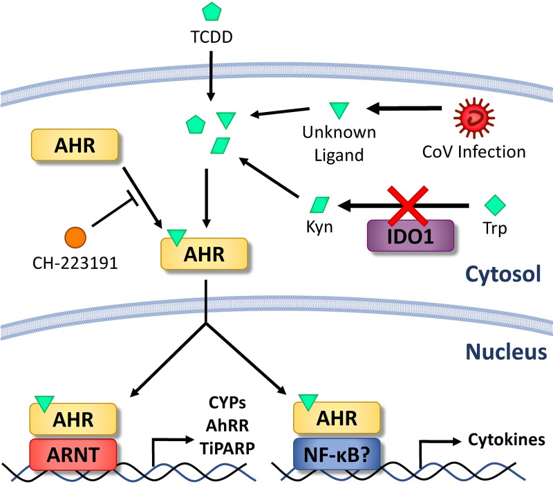
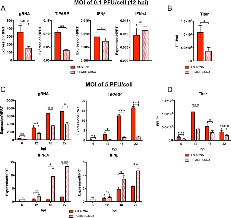
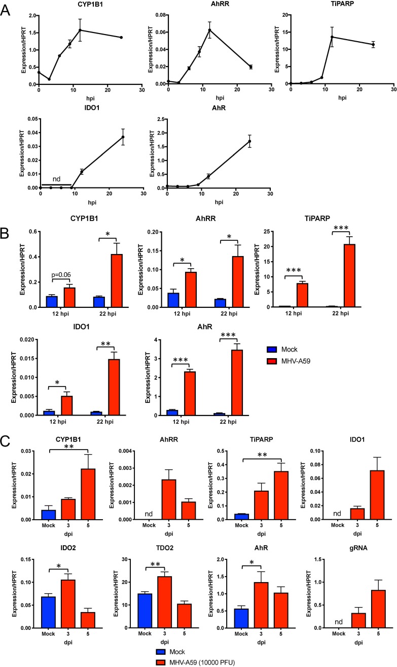
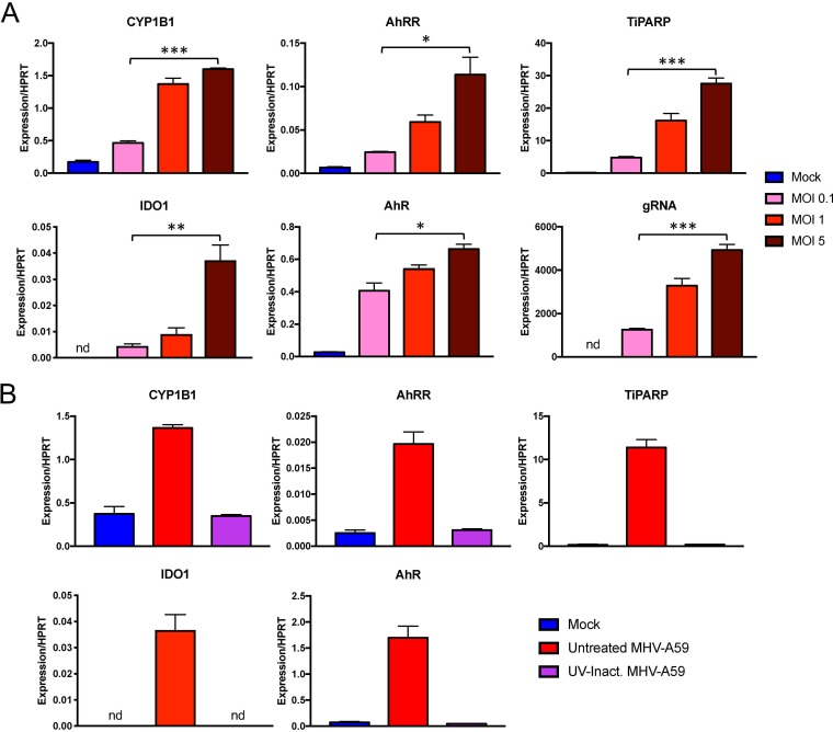
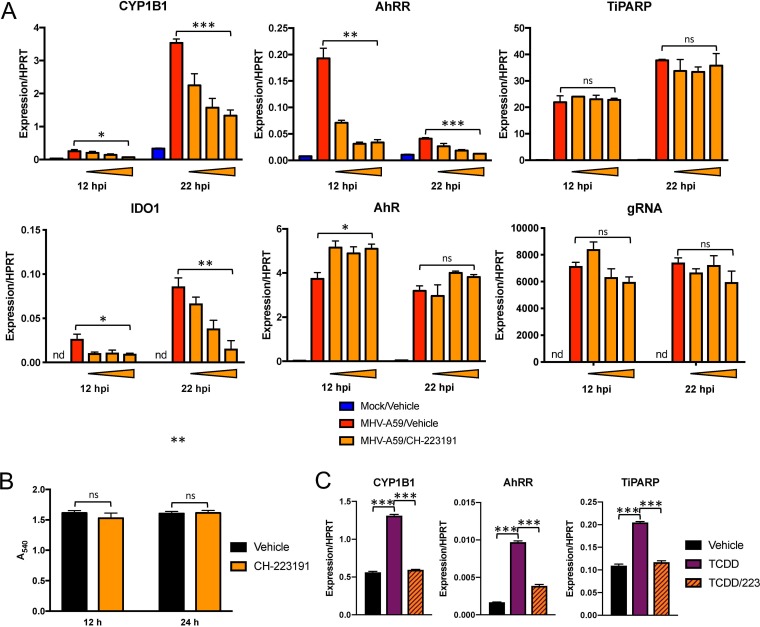
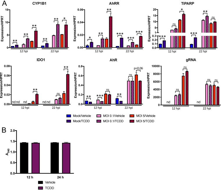
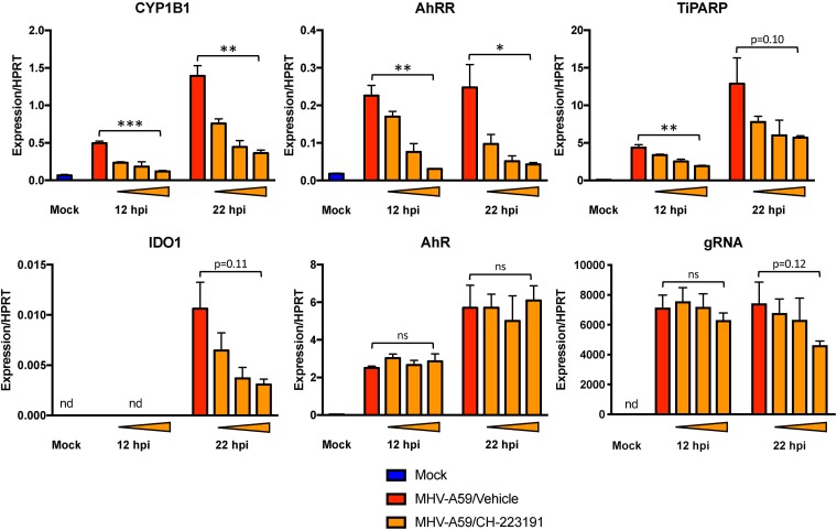
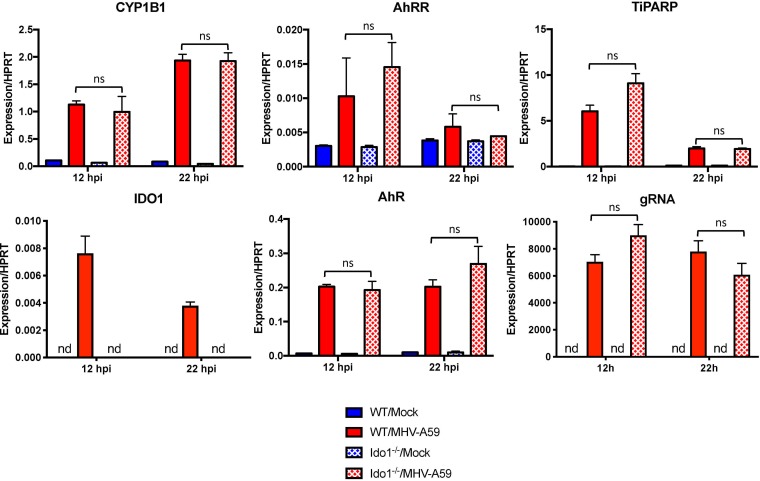
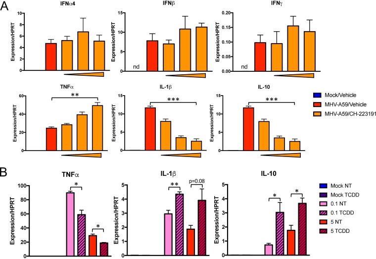
The aryl hydrocarbon receptor (AhR) is a cytoplasmic receptor/transcription factor that modulates several cellular and immunological processes following activation by pathogen-associated stimuli, though its role during virus infection is largely unknown. Here, we show that AhR is activated in cells infected with mouse hepatitis virus (MHV), a coronavirus (CoV), and contributes to the upregulation of downstream effector TCDD-inducible poly(ADP-ribose) polymerase (TiPARP) during infection. Knockdown of TiPARP reduced viral replication and increased interferon expression, suggesting that TiPARP functions in a proviral manner during MHV infection. We also show that MHV replication induced the expression of other genes known to be downstream of AhR in macrophages and dendritic cells and in livers of infected mice. Further, we found that chemically inhibiting or activating AhR reciprocally modulated the expression levels of cytokines induced by infection, specifically, interleukin 1β (IL-1β), IL-10, and tumor necrosis factor alpha (TNF-α), consistent with a role for AhR activation in the host response to MHV infection. Furthermore, while indoleamine 2,3-dioxygenase (IDO1) drives AhR activation in other settings, MHV infection induced equal expression of downstream genes in wild-type (WT) and IDO1-/- macrophages, suggesting an alternative pathway of AhR activation. In summary, we show that coronaviruses elicit AhR activation by an IDO1-independent pathway, contributing to upregulation of downstream effectors, including the proviral factor TiPARP, and to modulation of cytokine gene expression, and we identify a previously unappreciated role for AhR signaling in CoV pathogenesis.IMPORTANCE Coronaviruses are a family of positive-sense RNA viruses with human and agricultural significance. Characterizing the mechanisms by which coronavirus infection dictates pathogenesis or counters the host immune response would provide targets for the development of therapeutics. Here, we show that the aryl hydrocarbon receptor (AhR) is activated in cells infected with a prototypic coronavirus, mouse hepatitis virus (MHV), resulting in the expression of several effector genes. AhR is important for modulation of the host immune response to MHV and plays a role in the expression of TiPARP, which we show is required for maximal viral replication. Taken together, our findings highlight a previously unidentified role for AhR in regulating coronavirus replication and the immune response to the virus.

Keywords: AhR; IDO1; TiPARP; coronavirus; mouse hepatitis virus.


Figures

FIG 1
FIG 1
Schematic diagram of AhR activation in MHV-infected cells. Ligands that can bind to AhR can be exogenous (pentagons), such as toxins like TCDD, or endogenous metabolic products (parallelograms), such as kynurenine derived by IDO1-catalyzed tryptophan degradation. CoV infection produces an unknown AhR-activating ligand independent of IDO1 (triangles). AhR in the cytosol is activated upon ligand binding and translocates to the nucleus to bind to genomic DNA. The specific genes targeted and induced by AhR are influenced by AhR binding partners, including ARNT and NF-κB. Modulated genes include those encoding AhR downstream effectors, such as CYPs, AhRR, and TiPARP, or immune proteins, such as cytokines. AhR activation/ligand binding can be inhibited chemically by treatment with CH-223191.
FIG 2
FIG 2
MHV-A59 infection upregulates TiPARP in cell lines and primary cells in the absence of IFN-I signaling. (A and B) DBT (A) or 17Cl-1 (B) cell lines were infected with MHV-A59 at an MOI of 0.1 PFU/cell. Cells were collected at 18 hpi, and RNA was quantified by qRT-PCR with primers for the indicated PARPs. (C and D) WT (C) or WT and IFNAR−/− (D) BMDMs were mock infected or infected with MHV-A59 at an MOI of 5 (C) or 0.1 (D) PFU/cell. Cells were collected at 18 hpi, and mRNA was analyzed for quantification by qRT-PCR with primers for the indicated PARPs. The data show the results of one experiment representative of three independent experiments; n = 3. *, P ≤ 0.05; **, P ≤ 0.01; ***, P ≤ 0.001; ns, not significant; nd, not detectable. The data are expressed as means and SEM.
FIG 3
FIG 3
MHV replication is diminished following TiPARP knockdown in BMDMs. WT BMDMs were transfected with siRNA targeting TiPARP and were then infected at 30 h posttransfection with MHV-A59 at an MOI of 0.1 (A and B) or 5 (C and D) PFU/cell. RNA was quantified by qRT-PCR with primers specific for viral gRNA or for the indicated transcripts (A and C), or virus titers were determined by freezing-thawing cells, followed by plaque assay on HeLa cells expressing the MHV receptor (B and D). RNA and cell collection occurred at 12 hpi for an MOI of 0.1 PFU/cell (A and B) or at 6, 12, 18, and 22 hpi for an MOI of 5 PFU/cell (C and D). (A and C) The data show the results of one experiment representative of at least two independent experiments; n = 3. (B and D) The data show combined results of 2 experiments; n = 6. *, P ≤ 0.05; **, P ≤ 0.01; ***, P ≤ 0.001; ns, not significant. The data are expressed as means and SEM.
FIG 4
FIG 4
MHV infection results in upregulation of AhR downstream effector genes in BMDMs and BMDCs and in vivo. (A) BMDMs were infected with MHV-A59 at an MOI of 5 PFU/cell and collected at the indicated time points. mRNA levels of downstream effector AhR or IDO1 genes were then quantified by qRT-PCR. (B) BMDCs were infected with MHV-A59 at an MOI of 5 PFU/cell and collected at 12 or 22 hpi. mRNA levels of the indicated genes were quantified by qRT-PCR. (A and B) The data show the results of one experiment representative of two independent experiments; n = 3. (C) C57BL/6 mice were intraperitoneally infected with 104 PFU of MHV-A59, and perfused livers were harvested at 3 and 5 dpi. RNA was isolated, and mRNA levels of downstream effectors, AhR, or kynurenine-producing enzymes were quantified by qRT-PCR. The data show the results of one experiment representative of three independent experiments; n = 4. *, P ≤ 0.05; **, P ≤ 0.01; ***, P ≤ 0.001; nd, not detectable. The data are expressed as means and SEM.
FIG 5
FIG 5
Virus replication is correlated with and required for expression of AhR downstream effectors during infection. (A) BMDMs were infected with MHV-A59 at the indicated MOIs. Cells were then collected at 12 hpi, and mRNA levels of the downstream effectors AhR and IDO1 were quantified by qRT-PCR. (B) BMDMs were infected with untreated or UV-inactivated virus at an MOI of 5 PFU/cell. Expression of the indicated genes was quantified by qRT-PCR. The data show the results of one experiment representative of two independent experiments; n = 3. *, P ≤ 0.05; **, P ≤ 0.01; ***, P ≤ 0.001; nd, not detectable. The data are expressed as means and SEM.
FIG 6
FIG 6
Chemical inhibition of AhR during MHV infection results in attenuated expression of downstream genes. (A) WT BMDMs were pretreated with vehicle (0.01% DMSO) or 0.2, 1, or 5 μM CH-223191 and infected with MHV-A59 at an MOI of 5 PFU/cell. RNA was collected at 12 or 22 hpi and quantified for mRNA levels of the downstream effectors AhR and IDO1 and gRNA by qRT-PCR. The data show the results of one experiment representative of three independent experiments; n = 3. (B) Viability of BMDMs treated with vehicle or 5 μM CH-223191 was quantified by MTT assay at 12 and 24 h posttreatment. The data show the results of one experiment representative of two independent experiments; n = 4. (C) CH-223191 inhibitor efficacy was determined by preincubating uninfected BMDMs with vehicle (0.01% DMSO) or 5 μM CH-223191, followed by addition of vehicle or 10 nM TCDD. At 12 h, RNA was harvested, and mRNA levels of downstream effectors were determined by qRT-PCR. The data show the results of one experiment representative of two independent experiments; n = 3. *, P ≤ 0.05; **, P ≤ 0.01; ***, P ≤ 0.001; ns, not significant; nd, not detectable. The data are expressed as means and SEM.
FIG 7
FIG 7
Chemical activation of AhR in the absence and presence of infection enhances expression of downstream genes. (A) WT BMDMs were treated with vehicle (0.01% DMSO) or 10 nM TCDD and concurrently mock infected or infected with MHV-A59 at an MOI of 0.1 or 5 PFU/cell. RNA was collected at 12 or 22 hpi and quantified for mRNA levels of the downstream effectors AhR and IDO1 and gRNA by qRT-PCR. The data show the results of one experiment representative of two independent experiments; n = 3. (B) Viability of BMDMs treated with vehicle or 10 nM TCDD was quantified by MTT assay at 12 and 24 h posttreatment. The data show the results of one experiment representative of two independent experiments; n = 4. *, P ≤ 0.05; **, P ≤ 0.01; ***, P ≤ 0.001; ns, not significant; nd, not detectable. The data are expressed as means and SEM.
FIG 8
FIG 8
Infection upregulates TiPARP through redundant mechanisms requiring AhR activation or IFN-I signaling. BMDMs from IFNAR−/− mice were pretreated with vehicle (0.01% DMSO) or 0.2, 1, or 5 μM CH-223191 and infected with MHV-A59 at an MOI of 5 PFU/cell. RNA was collected at 12 or 22 hpi and quantified for mRNA levels of the downstream effectors AhR and IDO1 and gRNA by qRT-PCR. The data show the results of one experiment representative of two independent experiments; n = 3. *, P ≤ 0.05; **, P ≤ 0.01; ***, P ≤ 0.001; ns, not significant; nd, not detectable. The data are expressed as means and SEM.
FIG 9
FIG 9
IDO1 expression is dispensable for MHV-mediated AhR activation. BMDMs from WT or IDO1−/− mice were infected with MHV-A59 at an MOI of 5 PFU/cell. RNA was collected at 12 or 22 hpi, and expression of the indicated genes was quantified by qRT-PCR. The data show the results of one experiment representative of two independent experiments; n = 3. ns, not significant; nd, not detectable. The data are expressed as means and SEM.
FIG 10
FIG 10
Inhibition or enhancement of AhR activation during MHV infection modulates cytokine expression. (A) WT BMDMs were pretreated with vehicle (0.01% DMSO) or 0.2, 1, or 5 μM CH-223191 and then infected at an MOI of 5 PFU/cell. RNA was collected and quantified at 22 hpi for mRNA levels of the indicated cytokines by qRT-PCR. The data show the results of one experiment representative of three independent experiments; n = 3. (B) WT BMDMs were treated with vehicle (0.01% DMSO) or 10 nM TCDD and concurrently infected with MHV-A59 at an MOI of 0.1 or 5 PFU/cell. RNA was collected and quantified at 22 hpi for mRNA levels of the indicated cytokines by qRT-PCR. The data show the results of one experiment representative of two independent experiments; n = 3. *, P ≤ 0.05; **, P ≤ 0.01; ***, P ≤ 0.001; ns, not significant; nd, not detectable. The data are expressed as means and SEM.

References

    1. Fehr AR, Perlman S. 2015. Coronaviruses: an overview of their replication and pathogenesis. Methods Mol Biol 1282:1–23. doi: 10.1007/978-1-4939-2438-7_1. - DOI - PMC - PubMed
    1. Bergmann CC, Lane TE, Stohlman SA. 2006. Coronavirus infection of the central nervous system: host-virus stand-off. Nat Rev Microbiol 4:121–132. doi: 10.1038/nrmicro1343. - DOI - PMC - PubMed
    1. Roth-Cross JK, Bender SJ, Weiss SR. 2008. Murine coronavirus mouse hepatitis virus is recognized by MDA5 and induces type I interferon in brain macrophages/microglia. J Virol 82:9829–9838. doi: 10.1128/JVI.01199-08. - DOI - PMC - PubMed
    1. Ireland DD, Stohlman SA, Hinton DR, Atkinson R, Bergmann CC. 2008. Type I interferons are essential in controlling neurotropic coronavirus infection irrespective of functional CD8 T cells. J Virol 82:300–310. doi: 10.1128/JVI.01794-07. - DOI - PMC - PubMed
    1. Zhou H, Perlman S. 2006. Preferential infection of mature dendritic cells by mouse hepatitis virus strain JHM. J Virol 80:2506–2514. doi: 10.1128/JVI.80.5.2506-2514.2006. - DOI - PMC - PubMed

Publication types

MeSH terms

Substances

LinkOut - more resources