. 2020 Feb;39(7):1572-1589.
doi: 10.1038/s41388-019-1080-3. Epub 2019 Nov 6.

Nuclear receptor HNF4α performs a tumor suppressor function in prostate cancer via its induction of p21-driven cellular senescence

Affiliations

Nuclear receptor HNF4α performs a tumor suppressor function in prostate cancer via its induction of p21-driven cellular senescence

Zhu Wang et al. Oncogene. 2020 Feb.

Erratum in

Abstract

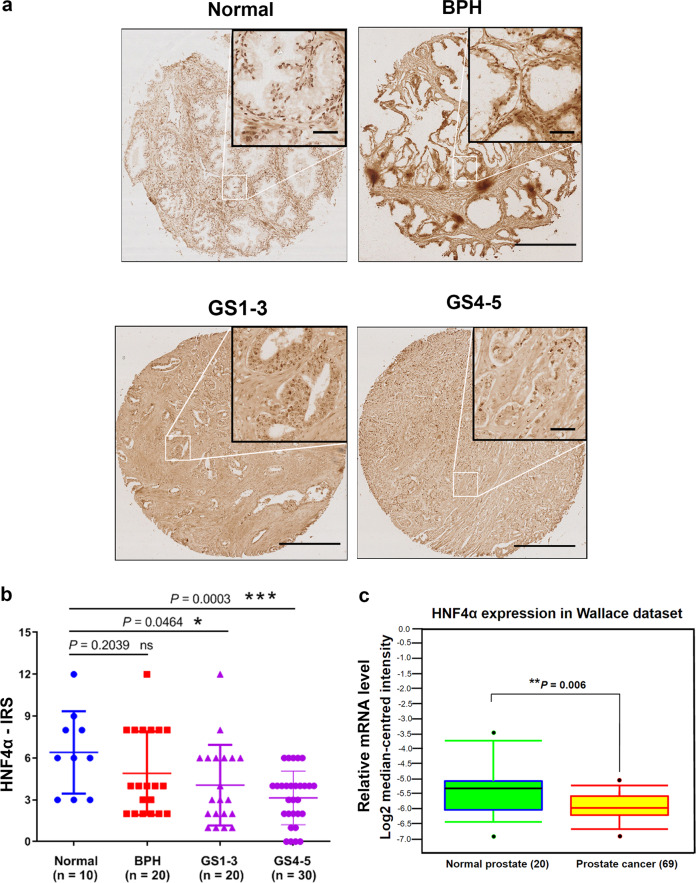
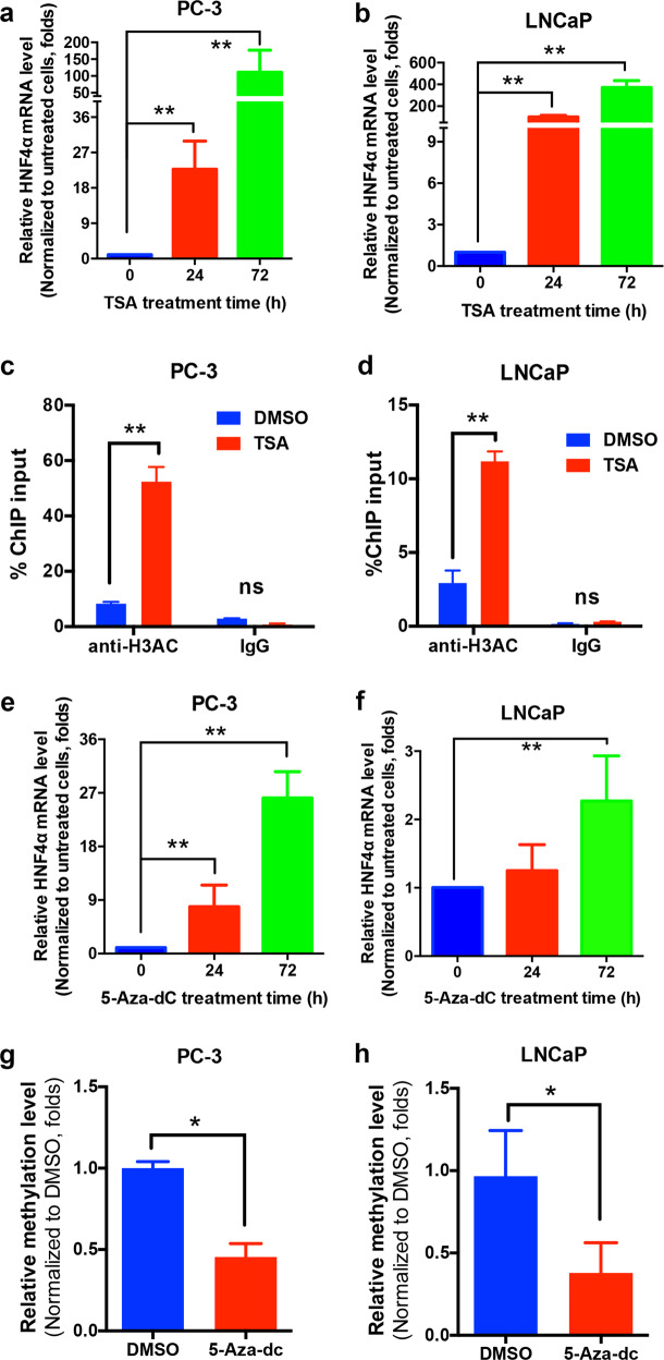
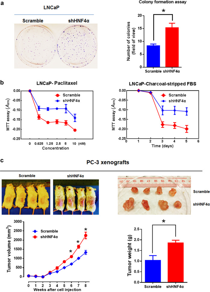
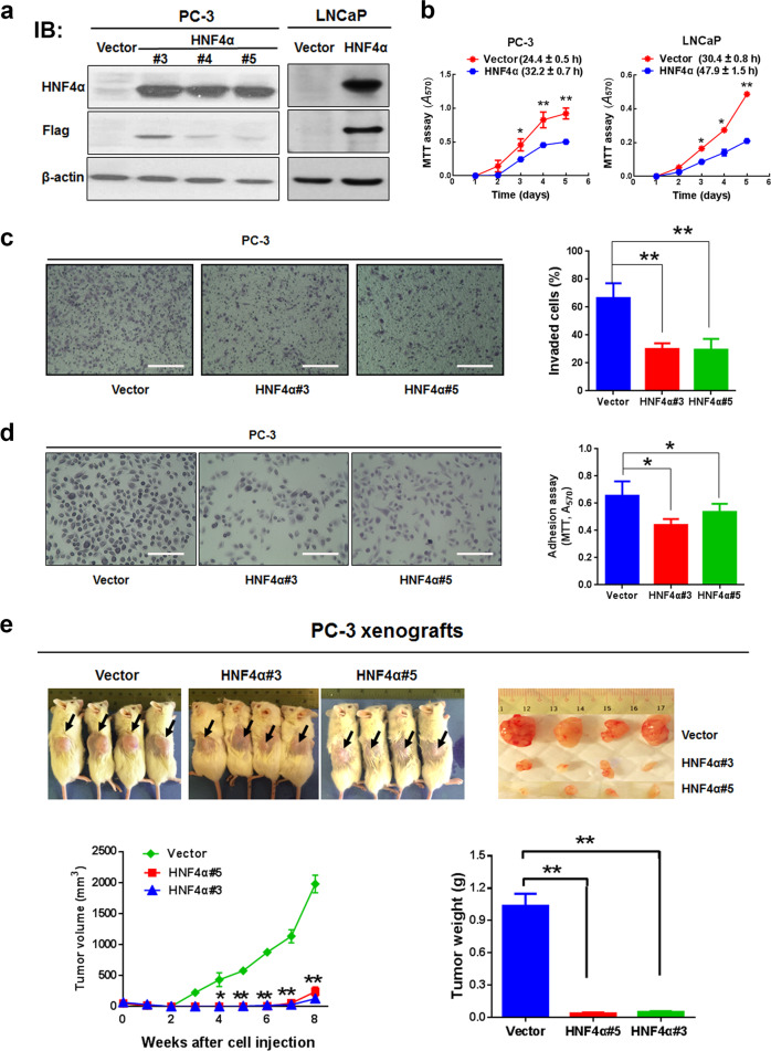
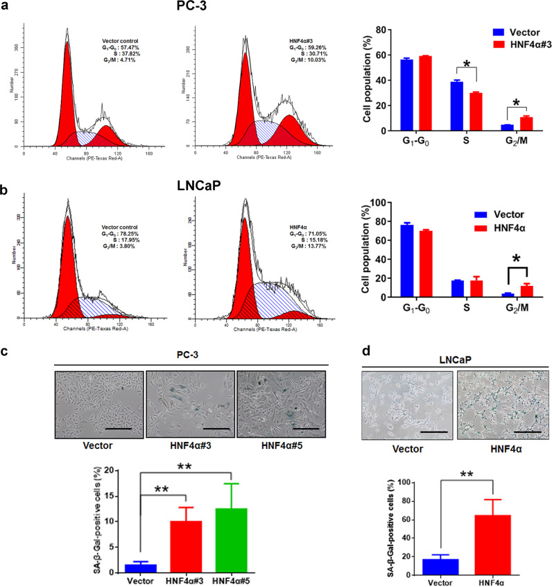
Hepatocyte nuclear factor 4α (HNF4α, NR2A1) is a highly conserved member of the nuclear receptor superfamily. Recent advances reveal that it is a key transcriptional regulator of genes, broadly involved in xenobiotic and drug metabolism and also cancers of gastrointestinal tract. However, the exact functional roles of HNF4α in prostate cancer progression are still not fully understood. In this study, we determined the functional significance of HNF4α in prostate cancer. Our results showed that HNF4α exhibited a reduced expression pattern in clinical prostate cancer tissues, prostate cancer cell lines and xenograft model of castration-relapse prostate cancer. Stable HNF4α knockdown not only could promote cell proliferation and suppress doxorubicin (Dox)-induced cellular senescence in prostate cancer cells, but also confer resistance to paclitaxel treatment and enhance colony formation capacity and in vivo tumorigenicity of prostate cancer cells. On the contrary, ectopic overexpression of HNF4α could significantly inhibit the cell proliferation of prostate cancer cells, induce cell-cycle arrest at G2/M phase and trigger the cellular senescence in prostate cancer cells by activation of p21 signal pathway in a p53-independent manner via its direct transactivation of CDKN1A. Together, our results show that HNF4α performs a tumor suppressor function in prostate cancer via a mechanism of p21-driven cellular senescence.


Conflict of interest statement

The authors declare that they have no conflict of interest.

Figures

Fig. 1
Fig. 1
HNF4α exhibits a decreased expression pattern in prostate cancer. a Immunohistochemical analysis of HNF4α in tissue microarray. Representative micrographs show the HNF4α-immunostained tissue spots of normal prostate, benign prostatic hyperplasia (BPH), and prostate cancer tissues. The normal prostatic and BPH epithelial cells exhibited moderate immunostaining of HNF4α in their nuclei. The malignant cells in low and moderate differentiated adenocarcinoma lesions (Gleason scores 1–3) showed weak nuclear immunoreactivity and barely detected or negative immunoreactivity in high-grade poorly differentiated lesions (Gleason scores 4–5). Magnification, ×40; scale bars = 500 μm. Insets show the enclosed areas at higher magnification. Magnification ×400; scale bars = 100 µm. b HNF4α-IRS analysis performed on malignant and nonmalignant (normal and BPH) prostatic tissues. Results showed that adenocarcinoma lesions exhibited significant lower HNF4α expression than that normal and BPH tissues. *P < 0.05, ***P < 0.001. IRS, immunoreactivity score. c Expression profile of HNF4α as revealed in an Oncomine dataset (GSE6956). Results showed that HNF4α mRNA levels exhibited a significant decrease in prostate cancer tissues as compared with normal prostate gland. Box plot (lines from top to bottom): maximum, third quartile Q3; median; minimum, first quartile Q1. **P < 0.01 versus normal prostate
Fig. 2
Fig. 2
Downregulation of HNF4α in prostate cancer cells involves epigenetic modifications. a, b qRT-PCR analysis of HNF4α expression. Results showed that HNF4α expression was significantly increased upon treatment with histone deacetylase inhibitor (TSA, 200 ng/ml) in both PC-3 (a) and LNCaP (b) cells. c, d ChIP-qPCR assay of HNF4A gene using H3Ac antibody (ab47915, abcam). Results validated that acetyl-histone H3 was highly enriched in the HNF4A promoter in both PC-3 (c) and LNCaP (d) cells. e, f qRT-PCR analysis. Results showed HNF4α expression was rescued in both PC-3 (e) and LNCaP (f) cells upon treatment with DNA methyltransferase inhibitor (5-aza-dC, 100 nM). g, h qMSP analysis. Results showed that there was a significant demethylation of the HNF4A gene promoter in PC-3 (g) and LNCaP (h) cells after 5-aza-dC treatment. *P < 0.05, **P < 0.01 versus untreated or DMSO group
Fig. 3
Fig. 3
HNF4α knockdown promotes cell proliferation and resistance to Dox-induced cellular senescence in prostate cancer cells. a Immunoblot and qRT-PCR validation of shRNA knockdown. Left: immunoblot validation of the constructed HNF4α-specific shRNA expressing vector on its HNF4α-knockdown efficiency in AR-negative PC-3 cells and AR-positive LNCaP cells. Right: qRT-PCR analysis showed that the HNF4α mRNA expression was significantly reduced in shHNF4α-infectants of PC-3 and LNCaP cells. b MTT analysis. Both PC-3-shHNF4α and LNCaP-shHNF4α infectants proliferated at faster rates than their shScramble infectants. c SA-β-Gal cytochemical assay. Top: micrographs show shHNF4α- and shScramble infectants upon Dox treatment (1 nM). Scale bars = 100 μm. Bottom: graphs show the % of SA-β-Gal-positive cells. Dox treatment induced no significant increase of SA-β-Gal-positive cells in shHNF4α-infectants as compared to shScramble infectants. *P < 0.05, **P < 0.01 versus shScramble infectants
Fig. 4
Fig. 4
HNF4α knockdown promotes both in vitro and in vivo malignant growth of prostate cancer cells. a Clonogenic assay. Left: representative images of stained colonies formed by the LNCaP-shHNF4α and LNCaP-shScramble infectants. Right: histogram shows the colonies formed by the LNCaP-shHNF4α and LNCaP-shScramble infectants. The LNCaP-shHNF4α infectants formed more and larger colonies than the LNCaP-shScramble infectants. b MTT assay. The LNCaP-shHNF4α infectants showed higher resistance to paclitaxel (0.625–10 nM) and charcoal-stripped FBS than LNCaP-shScramble infectants. c In vivo tumorigenicity assay. Upper panels: images of representative host SCID mice bearing the subcutaneous xenograft tumors formed by the injected PC-3-shHNF4α and PC-3-shScramble infectants (left) and the tumors dissected from the host mice (right). Lower panels: time-growth curve of xenograft tumors formed by the PC-3-shHNF4α and PC-3-shScramble infectants and the histogram of wet weights of tumors dissected from the host mice. The PC-3-shHNF4α infectants formed xenograft tumors at faster rates and with larger sizes than that of PC-3-shScramble infectants. *P < 0.05 versus shScramble infectants
Fig. 5
Fig. 5
HNF4α overexpression suppresses both in vitro and in vivo malignant growth of prostate cancer cells. a Immunoblot validation of HNF4α protein expression in stable HNF4α-infectants generated from PC-3 and LNCaP cells. Vector-infectants were set as controls and detection of Flag-tagged HNF4α was used as positive expression control. b Cell viability analysis. The growth curves of HNF4α-overexpressed PC-3 (left) and LNCaP (right) cells were determined by MTT assay. Results showed that HNF4α-infectants grew significantly slower than their corresponding vector-infectants. Brackets show the doubling time (h) of infectants. c In vitro invasion assay. Left: representative micrographs show the crystal violet-stained invaded cells on membranes. Scale bars = 100 μm. Right: graph shows the number of invaded cells counted in five randomly selected ×200 fields per transwell insert. All determinations were performed at least in triplicate in three independent experiments. d Adhesion assay. Left: representative micrographs show the crystal violet-stained adherent cells. Scale bars = 100 μm. Right: graph shows the adherent cell number as determined by MTT assay. Results showed that there was a significant decrease of adherent cells in HNF4α overexpressed PC-3 cells. All determinations were performed at least in triplicate in three independent experiments. e In vivo tumorigenicity assay. Upper: representative images of mice bearing xenograft tumors and dissected tumors formed by HNF4α- or vector-infectants. Lower: time-growth curve of xenograft tumors grown in mice and histogram shows the wet weights of dissected tumors. All determinations were performed at least in triplicate in three independent experiments. The data are presented as the mean ± SD from three independent experiments. *P < 0.05; **P < 0.01 versus vector-infectant controls
Fig. 6
Fig. 6
HNF4α overexpression induces cell-cycle arrest and cellular senescence in prostate cancer cells. a, b Cell cycle analysis. Flow cytometric analysis of PC-3 and LNCaP-HNF4α-infectants and their vector-infectants control shown as DNA histograms. Cell-cycle distribution was represented as the percentage of cells at each phase. Both PC-3 and LNCaP-HNF4α-infectants showed significant higher percentages of cells at G2/M phase than their control vector-infectants. c, d Cytochemical staining of SA-β-Gal activity in HNF4α-infectants. Upper: representative micrographs show the SA-β-Gal staining (perinuclear staining) in HNF4α-infectants. Scale bars = 100 μm. Lower: histograms show the percentages of SA-β-Gal-positive cells in HNF4α-infectants. Results showed that there was a significant increase of SA-β-Gal-positive cells in both PC-3 and LNCaP-HNF4α-infectants as compared with their control vector-infectants. *P < 0.05; **P < 0.01 versus control vector-infectants
Fig. 7
Fig. 7
Knockdown of HNF4α prevents cellular senescence induced by activated oncogene H-RasG12V or loss-of-PTEN in prostatic epithelial cells. Evaluation of SA-β-Gal activity in RasG12V-transduced and shPTEN-transduced (a) PrEC and (b) PWR-1E cells upon HNF4α knockdown. Upper: representative micrographs show the SA-β-Gal-positive cells detected in RasG12V-transduced (left panels) and shPTEN-transduced (right panels) PrEC and PWR-1E cells with or without HNF4α knockdown. Lower: histograms show the scores of SA-β-Gal-positive cells in RasG12V and shPTEN-transduced PrEC and PWR-1E cells with or without HNF4α knockdown. Magnification, × 200; scale bars = 100 μm. *P < 0.05; **P < 0.01 versus no shHNF4α infection
Fig. 8
Fig. 8
HNF4α-induced cellular senescence in prostate cancer cells is driven by its direct transactivation of p21 (CDKN1A) gene. a qRT-PCR (left) and immunoblot (right) analyses of p21 expression in HNF4α-overexpressed or HNF4α-silenced PC-3 cells. Results showed that both p21 mRNA and protein levels were significantly increased in PC-3-HNF4α infectants, but decreased in shHNF4α-PC-3 infectants. b Detection and scoring of SA-β-Gal-positive cells in PC-3-HNF4α infectants upon p21 knockdown. Upper: representative micrographs show the SA-β-Gal-positive cells in PC-3-HNF4α infectants with or without p21 knockdown. Lower: histogram shows the SA-β-Gal-positive cells. Results showed that knockdown of p21 could significantly reduce the number of SA-β-Gal-positive cells in PC-3-HNF4α infectants. Results were obtained from three independent experiments. Scale bars = 100 μm. c Luciferase reporter assay. Left: luciferase reporter assay of CDKN1A-Luc reporter performed in HEK293 cells transfected with either intact HNF4α or its truncated mutants (HNF4α-ΔDBD or HNF4α-ΔLBD). Results showed that only the intact HNF4α but not its truncated mutants could transactivate the CDKN1A-Luc reporter in a dose-dependent manner. Right: luciferase reporter assay performed in PC-3-HNF4α infectants transfected with either CDKN1A-Luc or empty reporter pGL3. Results showed that significant transactivation of CDKN1A-Luc reporter was shown in PC-3-HNF4α infectants. *P < 0.05; **P < 0.01 versus controls. d ChIP assay. Top: schematic diagram shows the locations of two identified HNF4α-binding sites (P1, P2) located at the proximal region of CDKN1A gene promoter. The sequences of HNF4α-binding sites are shown in red. The locations of ChIP-PCR primers (indicated by arrows) are also shown. Bottom: results showed that only HNF4α but not IgG-immunoprecipitated DNA fragments extracted from HNF4α-transfected HEK293 cells could be PCR-amplified at the HNF4α-binding sites. Nonimmunoprecipitated sonicated DNA (10% diluted) was used as input. e Schematic diagram depicts the hypothesized mechanism of HNF4α-induced cellular senescence and growth arrest via its transactivation of p21 in prostate cancer cells

References

    1. Loaiza N, Demaria M. Cellular senescence and tumor promotion: Is aging the key? Biochim Biophys Acta. 2016;1865:155–67. - PubMed
    1. Muller M. Cellular senescence: Molecular mechanisms, in vivo significance, and redox considerations. Antioxid Redox Signal. 2009;11:59–98. - PubMed
    1. Sharma S, Shin JS, Grimshaw M, Clarke RA, Lee CS. The senescence pathway in prostatic carcinogenesis. Pathology. 2010;42:507–11. - PubMed
    1. Attard G, Parker C, Eeles RA, Schroder F, Tomlins SA, Tannock I, et al. Prostate cancer. Lancet. 2016;387:70–82. - PubMed
    1. Moschini M, Carroll PR, Eggener SE, Epstein JI, Graefen M, Montironi R, et al. Low-risk prostate cancer: Identification, management, and outcomes. Eur Urol. 2017;72:238–49. - PubMed

Publication types

MeSH terms

Substances