. 2019 Nov;26(11):1053-1062.
doi: 10.1038/s41594-019-0324-9. Epub 2019 Nov 6.

UPR proteins IRE1 and PERK switch BiP from chaperone to ER stress sensor

Affiliations

UPR proteins IRE1 and PERK switch BiP from chaperone to ER stress sensor

Megan C Kopp et al. Nat Struct Mol Biol. 2019 Nov.

Abstract

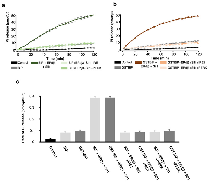
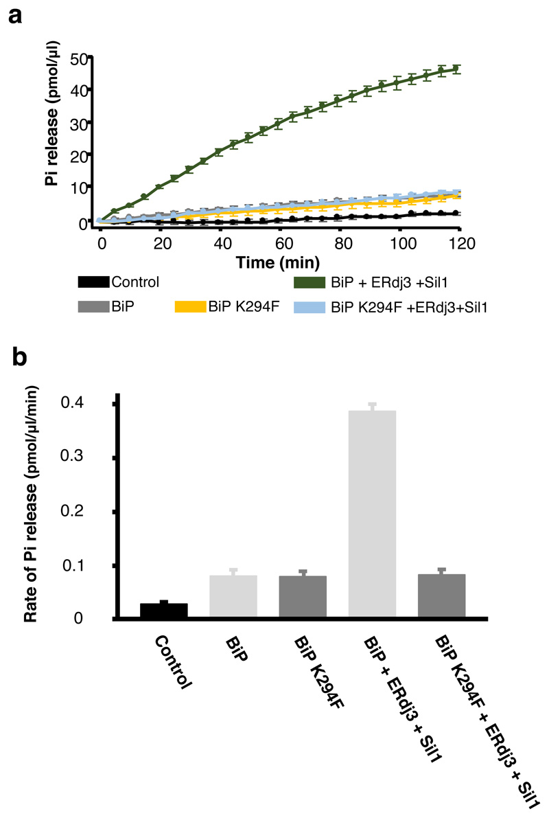
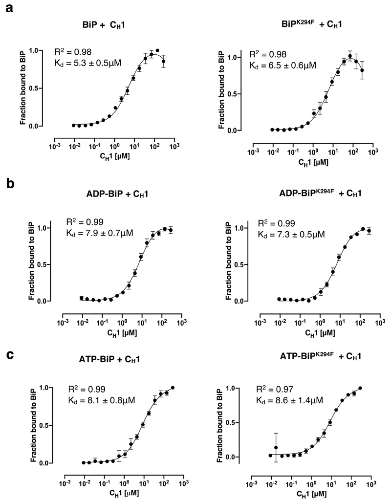
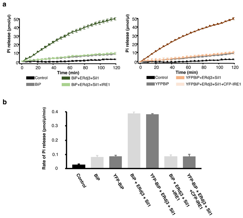
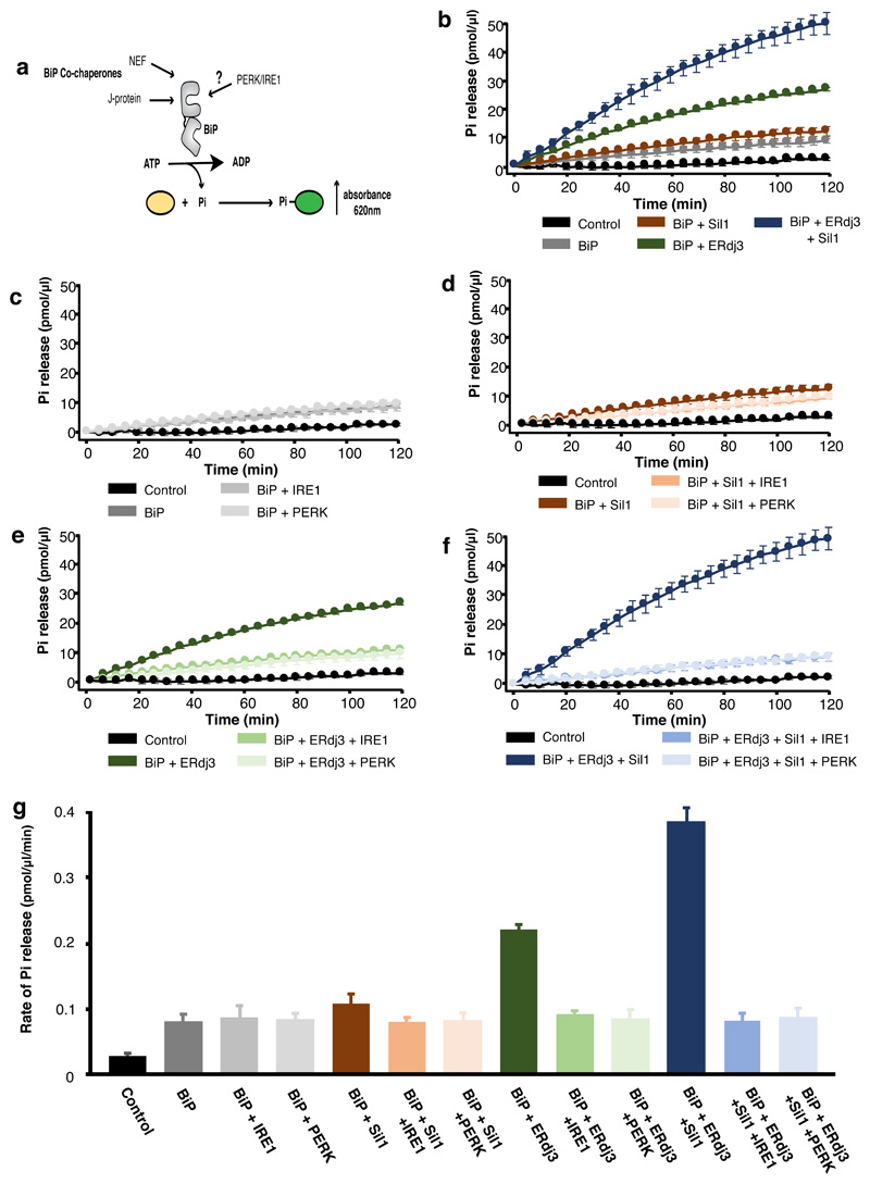
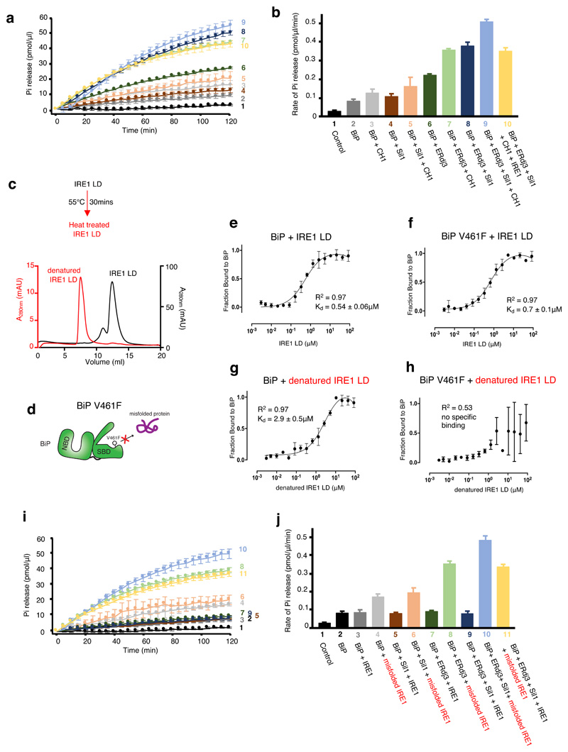
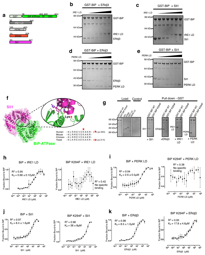
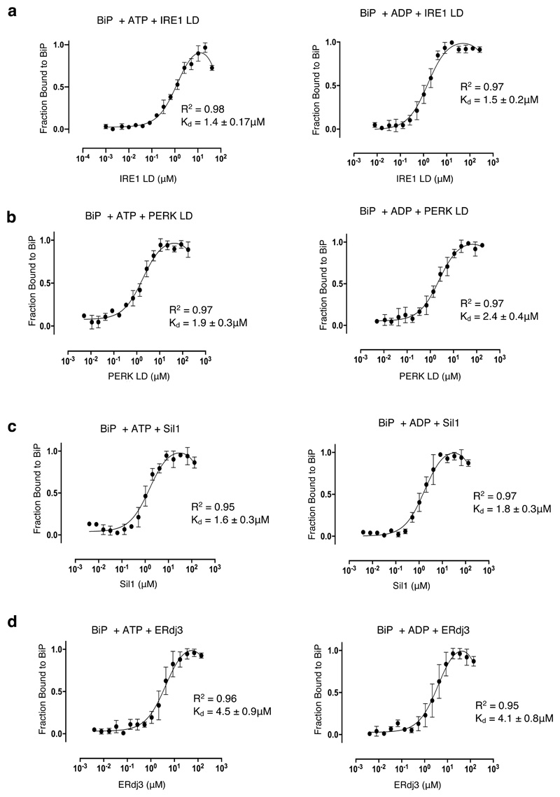
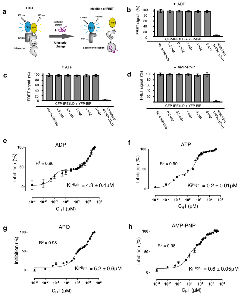
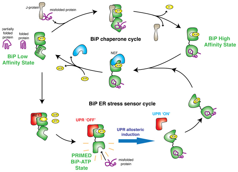
BiP is a major endoplasmic reticulum (ER) chaperone and is suggested to act as primary sensor in the activation of the unfolded protein response (UPR). How BiP operates as a molecular chaperone and as an ER stress sensor is unknown. Here, by reconstituting components of human UPR, ER stress and BiP chaperone systems, we discover that the interaction of BiP with the luminal domains of UPR proteins IRE1 and PERK switch BiP from its chaperone cycle into an ER stress sensor cycle by preventing the binding of its co-chaperones, with loss of ATPase stimulation. Furthermore, misfolded protein-dependent dissociation of BiP from IRE1 is primed by ATP but not ADP. Our data elucidate a previously unidentified mechanistic cycle of BiP function that explains its ability to act as an Hsp70 chaperone and ER stress sensor.


Conflict of interest statement

Competing interests

The authors declare no competing interests.

Figures

Extended Data Fig. 1
Extended Data Fig. 1. Comparison of BiP and GST-BiP stimulation and inhibition.
(a) BiP ATPase activity showing stimulation by co-chaperones and inhibition by IRE1 and PERK LD. (b) same as (a), but using GST tagged BiP. (c) A comparison of the rate of ATPase activity for BiP and GST tagged BiP. Both tagged and untagged BiP are stimulated by co-chaperones and inhibited by IRE1 and PERK LD to the same level, indicating that the attachment of GST to BiP had no effect on BiP ATPase stimulation or inhibition. Statistics as in Figure 1, source data available online.
Extended Data Fig. 2
Extended Data Fig. 2. Mutant BiPK294F has same basal ATPase activity as BiPWT.
(a) BiPK294F ATPase activity on addition of co-chaperones. (b) A comparison of the rate of ATPase activity for BiP and BiPK294F. The K294F mutation based within the BiP NBD, had no effect on inherent BiP ATPase, but prevents ATPase stimulation consistent with-it inhibiting co-chaperone binding. Statistics as in Figure 1, source data available online.
Extended Data Fig. 3
Extended Data Fig. 3. Assessment of BiPK294F affinity for CH1 in presence of nucleotides.
BiPK294F has same binding affinity for CH1, in different nucleotide bound states, as BiPWT. (a) MST profile measuring the binding affinity between BiP and BiPK294F for CH1. (b) same as (a), but in the presence of ADP. (C) same as (a), but with ATP. The experiments indicate that the mutation had no effect on BiP interaction with misfolded substrate protein or affected BiP nucleotide bound conformations. Statistics as in Figure 4, source data available online.
Extended Data Fig. 4
Extended Data Fig. 4. Attachment of YFP had no effect on BiP functionality
(a) ATPase activity of BiP and YFP tagged BiP. (b) Comparison of the rate of ATPase activity for BiP and YFP- tagged BiP on addition of co-chaperones and CFP-IRE1 LD. The attachment of YFP had no effect on BiP basal activity, or stimulation by co-chaperones, or inhibition by CFP tagged IRE1 LD. Statistics as in Figure 1, source data available online.
Figure 1
Figure 1. IRE1 and PERK LD cause loss of BiP ATPase stimulation by cochaperone and NEF effectors, but BiP retains its inherent activity.
a, Schematic representation of assay used for measuring ATPase activity. b-f, Graphs displaying phosphate (Pi) release over time as a result of BiP ATPase activity. Data shown are mean ± sd, from nine independent experiments. Source data available online. BiP displayed low basal ATPase activity; the addition of Sil1 slightly increased activity, whereas ERdj3 or both cochaperone Sil1 and ERdj3 together greatly stimulated BiP activity (b). Addition of IRE1 LD or PERK LD did not affect BiP basal ATPase activity (c) but reversed the stimulation by Sil1 (d), by ERdj3 (e), and by both cochaperones ERdj3 and Sil1 together (f). g, Bar graph showing the rates of BiP ATPase activity upon addition of effector proteins. Data are mean ± sd, from nine independent experiments. ATPase rates are in Supplementary Table 1.
Figure 2
Figure 2. Folded and misfolded IRE1 LD have different effects on BiP interaction and activity.
a-b, ATPase assay that measured BiP activity on addition of CH1 misfolded protein with cochaperones and IRE1 LD. Pi release over time (a) and rate of BiP ATPase activity (b) deduced from a. Data are mean ± sd from nine independent experiments. c, Size exclusion chromatography of IRE1 LD heat treated (55°C for 30 mins, red) or untreated control (black). The heat-treated sample eluted in the void volume indicating protein aggregation. d, A graphical representation of BiP V461F mutant. The SBD mutant prevents the engagement of misfolded chaperone-substrate protein to BiP,. e-f, Microscale thermophoresis (MST) profile of folded IRE1 LD interaction with BiPWT (e) or BiPV461F(f). The binding affinities are comparable and suggest folded IRE1 LD does not engage the SBD as a chaperone substrate. g-h, MST profile of misfolded IRE1 LD interaction with BiPWT (g), and BiPV461F(h). Data points in e-h are mean ± sd from three independent experiments. i, BiP ATPase assay comparing the effects of folded and misfolded IRE1 on BiP activity in the presence of cochaperones. j, rate of BiP ATP activity deduced from i (mean ± sd, n=9 independent experiments). Source data for graphs are available online. ATPase rate values for b and j are listed in Supplementary Tables 2 and 4. Binding affinities are in Supplementary Table 3.
Figure 3
Figure 3. UPR proteins and cochaperones have mutually exclusive binding sites on BiP NBD that are impacted by BiPK294F mutant.
a, Schematic representation of the various constructs used for the pull-down assay, with the numbers denoting the residue number of the protein. b, Competitive pull-down assay in which increasing concentrations of IRE1 LD were added to GST-BiP – ERdj3 chaperone complex. IRE1 LD outcompeted ERdj3 for binding to BiP, suggesting mutually exclusive binding. c, Competitive pull-down assay where GST-BiP - Sil1 complex was immobilized to beads and increasing concentrations of IRE1 LD were added. d, Same as (b), but with PERK LD. e, Same as (c), but with PERK LD. f, Crystal structure of BiP ATPase – Sil1 complex (PDB 3QML), highlighting the interface between interacting molecules. BiP NBD domain residue Lys314 (corresponding to Lys294 in human) mediates key polar contacts between the two molecules. Sequence alignment (right) indicated that this residue is highly conserved across BiP species. g, Pull-down assay to assess interaction of GST- BiPWT or BiPK294F mutant with cochaperones and UPR proteins IRE1 LD and PERK LD. h-k, MST experiments comparing the binding affinity of GST tagged BiPWT and BiPK294F to cochaperones and UPR proteins. Data are mean ± sd from 3 independent experiments. Source Data are available online. BiPK294F mutant disrupted binding to IRE1 LD (h) and PERK LD (i). The BiPK294F mutant displayed greatly reduced binding affinity for Sil1 compared to BiPWT (j). The BiPK294F mutant displayed reduced, but still substantial binding for ERdj3 indicating that ERdj3 may have other contact sites with BiP. Uncropped gel images are available as Source Data. Binding affinities are in Supplementary Table 5.
Figure 4
Figure 4. The effect of nucleotides on BiP binding to UPR proteins and cochaperones.
MST profiles measuring the binding between GST tagged BiP with UPR proteins and with cochaperones in the presence of ATP and ADP. a, The binding affinities for BiP association to IRE1 LD in the presence of ATP and ADP are similar to the binding affinity without nucleotides (Figure 3h). b, PERK LD incubated with ATP and ADP are similar to the binding affinity without nucleotides (Figure 3i). c-d, Sil1 (c) and ERdj3 (d) both show stronger binding to BiP with nucleotide present than without nucleotides (Figure 3 j-k). Data are mean ± sd from 3 independent experiments. Source Data are available online. Binding affinities are in Supplementary Table 6.
Figure 5
Figure 5. ATP – but not ADP – primes and facilitates the release of BiP from IRE1 LD.
a, Experimental design for three-component in vitro UPR FRET assay, used to measure the inhibition of CFP-IRE1 – YFP-BiP complex by misfolded protein in the presence and absence of nucleotides. b-d, Bar graph showing FRET signal observed upon addition of ADP (b), ATP (c) or AMP-PNP (d). In each case, FRET inhibition due to dissociation of the complex was solely dependent on binding of misfolded protein CH1. Data are mean ± s.d., n=9 independent experiments. e-h, FRET signal inhibition (%) as a function of the CH1 concentration in the absence and presence of different nucleotides: ADP (e), ATP (f), APO (g) and AMP-PNP (h). Data were fitted to exponential curve (two phase association) and the IC50 (equivalent to half-life) for each phase was deduced. The IC50 measurement was then used to calculate the Ki. The ATP bound BiP-IRE1 LD complex required 21-fold less misfolded protein to facilitate full dissociation of complex than the ADP bound state, suggesting a priming effect towards misfolded protein on ATP addition. Data are mean ± s.d., n = 9 independent experiments. Source Data are available online. The IC50 and inhibition constant values for both low and high concentration phases in the absence and presence of different nucleotides are in Supplementary Table 7.
Figure 6
Figure 6. Mechanism of BiP function encompassing its chaperone and ER stress sensor cycles.
Interaction with UPR proteins switch BiP from chaperone cycle to ER stress sensor cycle by preventing binding to cochaperone and NEF proteins, with loss of BiP ATPase stimulation. The interaction between IRE1 LD is mediated via BiP NBD, whilst dissociation of complex is dependent on misfolded protein binding to BiP SBD. The BiP-IRE1 complex in the ATP bound state, has a greater sensitivity for misfolded protein, thereby facilitating release. By contrast the complex is desensitized to misfolded protein when bound to ADP. The release of BiP from UPR proteins via conformational change, is coupled to UPR activation, enabling BiP to interact with cochaperones and revert to its chaperone cycle with misfolded protein attached.

References

    1. Hetz C, Papa FR. The Unfolded Protein Response and Cell Fate Control. Mol Cell. 2017 - PubMed
    1. Wang M, Kaufman RJ. Protein misfolding in the endoplasmic reticulum as a conduit to human disease. Nature. 2016;529:326–335. - PubMed
    1. Walter P, Ron D. The unfolded protein response: from stress pathway to homeostatic regulation. Science. 2011;334:1081–1086. - PubMed
    1. Adams CJ, Kopp MC, Larburu N, Nowak PR, Ali MMU. Structure and Molecular Mechanism of ER Stress Signaling by the Unfolded Protein Response Signal Activator IRE1. Front Mol Biosci. 2019;6:11. - PMC - PubMed
    1. Zhou J, et al. The crystal structure of human IRE1 luminal domain reveals a conserved dimerization interface required for activation of the unfolded protein response. Proc Natl Acad Sci USA. 2006;103:14343–14348. - PMC - PubMed

Publication types

MeSH terms