. 2019 Aug 27:13:1641-1643.
doi: 10.2147/OPTH.S221197. eCollection 2019.

Three-dimensional ocular endoscope system for vitrectomy

Affiliations

Three-dimensional ocular endoscope system for vitrectomy

Mihori Kita et al. Clin Ophthalmol. .

Abstract

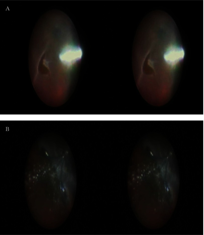
Purpose: To introduce a new three-dimensional (3D) endoscope system for vitrectomy.

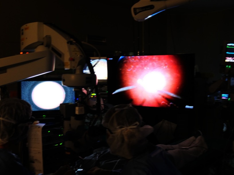
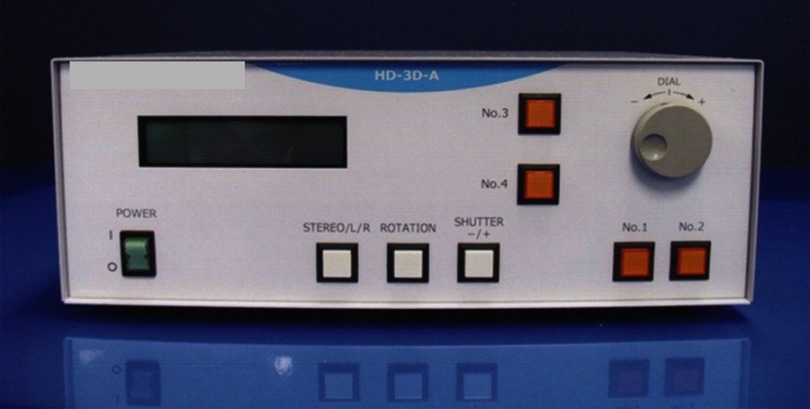
Subjects and methods: We report a single-center, retrospective, consecutive surgical case series of 391 eyes that underwent 25G hybrid vitrectomy. To create 3D endoscopic images, a 3D converter HD-3D-A was connected to a monocular endoscopic system.

Results: The 3D endoscope system was successfully used to perform hybrid vitrectomy. No intra- or postoperative complications related to this system were encountered in any of the cases.

Conclusion: This 3D endoscope system appears to be a valuable and promising tool for use in vitrectomy.

Keywords: 3D; endoscope; hybrid; vitrectomy; wide-angle view.


Conflict of interest statement

The authors report no conflicts of interest in this work.

Figures

Figure 1
Figure 1
The wide-angle viewing image is integrated on the large NGENUITY 3D monitor (right) that is placed at the foot of the surgical bed. The 3D monitor for the endoscope (left) is placed 1 m to the front left of the surgeon. Abbreviation: 3D, three-dimensional.
Figure 2
Figure 2
A 3D converter NOVEL HD-3D-A creates 3D endoscopic images in real-time. Abbreviation: 3D, three-dimensional.
Figure 3
Figure 3
Intraoperative processed 3D endoscope images are recorded as side-by-side stereo images. Notes: (A) Peripheral vitreous shaving around a retinal break was performed while using the endoscopic view without any scleral depression. (B) Silicone oil was removed through the cannula. Emulsified silicone oil was remarkable. Abbreviation: 3D, three-dimensional.

References

    1. Kita M, Yoshimura N. Endoscope-assisted vitrectomy in the management of pseudophakic and aphakic retinal detachments with undetected retinal breaks. Retina. 2011;31:1347–1351. doi:10.1097/IAE.0b013e3182003c93 - DOI - PubMed
    1. Yoshitake S, Oh H, Kita M. Endoscope-assisted vitrectomy for retinal detachment in an eye with microcornea. Jpn J Ophthalmol. 2012;56:613–616. doi:10.1007/s10384-012-0176-x - DOI - PubMed
    1. Kita M. Endoscope-assisted vitrectomy. World J of Ophthalmol. 2014;12:52–55. doi:10.5318/wjo.v4.i3.52 - DOI
    1. Kita M, Fujii Y, Hama S. Twenty five-gauge endoscopic vitrectomy for proliferative vitreoretinopathy with severe corneal opacity. Jpn J Ophthalmol. 2018;62:302–306. doi:10.1007/s10384-018-0578-5 - DOI - PubMed
    1. Morishita S, Kita M, Yoshitake S, Hirose M, Oh H. 23-gauge vitrectomy assisted by combined endoscopy and a wide-angle viewing system for retinal detachment with severe penetrating corneal injury: a case report. Clin Ophthalmol. 2011;5:1767–1770. doi:10.2147/OPTH.S25373 - DOI - PMC - PubMed

LinkOut - more resources