

Intramural Esophageal Hematoma Secondary to Food Ingestion
- PMID: 31696016
- PMCID: PMC6820886
- DOI: 10.7759/cureus.5623
Intramural Esophageal Hematoma Secondary to Food Ingestion
Abstract
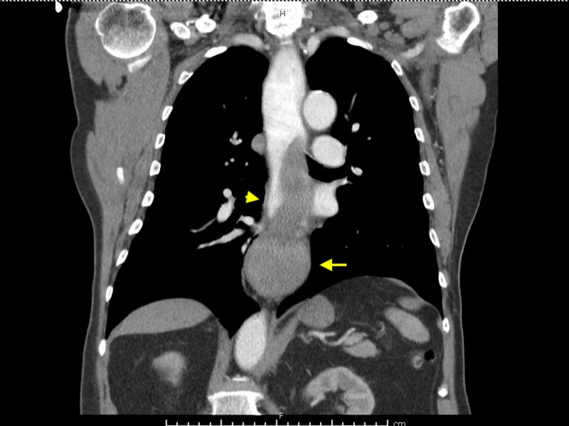
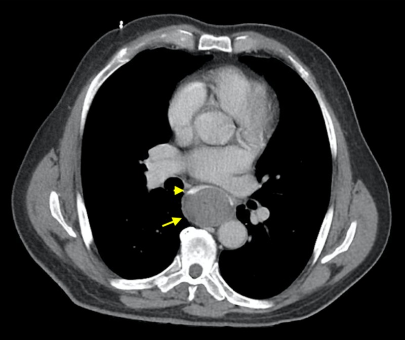
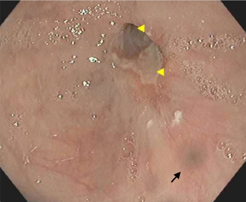
Intramural esophageal hematoma (IEH) is a rare cause of submucosal esophageal bleeding and it is on the spectrum of esophageal wall injury along with mucosal tears (Mallory-Weiss syndrome) and full thickness perforation (Boerhaave's syndrome). Its risk factors include coagulopathy, trauma (foreign body ingestion or esophageal instrumentation) or it can happen spontaneously. It presents with a triad of chest pain, dysphagia, and hematemesis; however, the triad is only present in 35% of patients. We are presenting a case of IEH secondary to food ingestion that was managed successfully by conservative measures.
Keywords: esophageal hematoma; esophageal wall injury; food ingestion.
Copyright © 2019, Sharma et al.
Conflict of interest statement
The authors have declared that no competing interests exist.
Figures
References
-
- The spectrum of spontaneous and iatrogenic esophageal injury: perforations, Mallory-Weiss tears, and hematomas. Younes Z, Johnson DA. https://www.ncbi.nlm.nih.gov/pubmed/10599632. J Clin Gastroenterol. 1999;29:306–317. - PubMed
-
- Intramural rupture and intramural hematoma of the esophagus: 3 case reports and literature review [Article in German] Criblez D, Filippini L, Schoch O, Meier UR, Koelz HR. https://www.ncbi.nlm.nih.gov/pubmed/1557620. Schweiz Med Wochenschr. 1992;122:416–423. - PubMed
-
- Intramural haematoma of the oesophagus complicating sclerotherapy for varices. Mosimann F, Brönnimann B. https://www.ncbi.nlm.nih.gov/pubmed/8307434. Gut. 1994;35:130–131. - PMC - PubMed
-
- A rare cause of melena: spontaneous esophageal hematoma. Chen HL, Chang WH, Shih SC, Lin SC, Wang TE, Chu CH. Gastrointest Endosc. 2008;67:539. - PubMed
Publication types
LinkOut - more resources
Full Text Sources