Review
. 2019 Dec 1;127(6):1632-1639.
doi: 10.1152/japplphysiol.00624.2019. Epub 2019 Nov 7.

Fiber typing human skeletal muscle with fluorescent immunohistochemistry

Affiliations
Review

Fiber typing human skeletal muscle with fluorescent immunohistochemistry

Kevin A Murach et al. J Appl Physiol (1985). .

Abstract

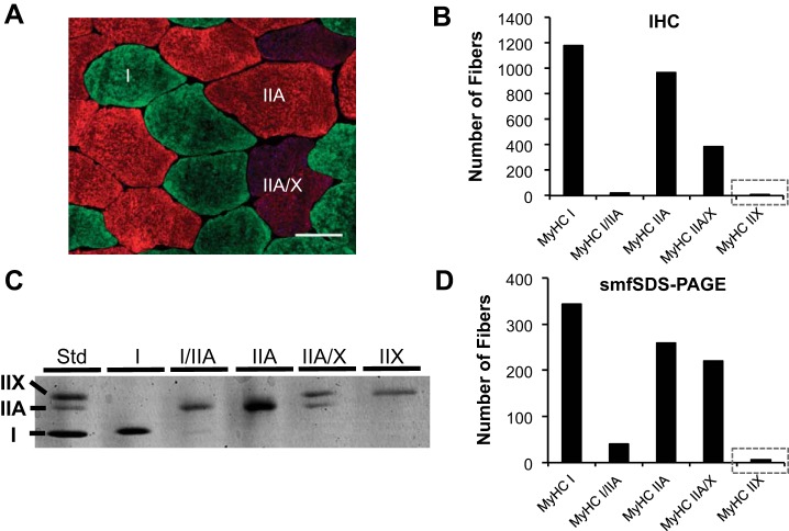
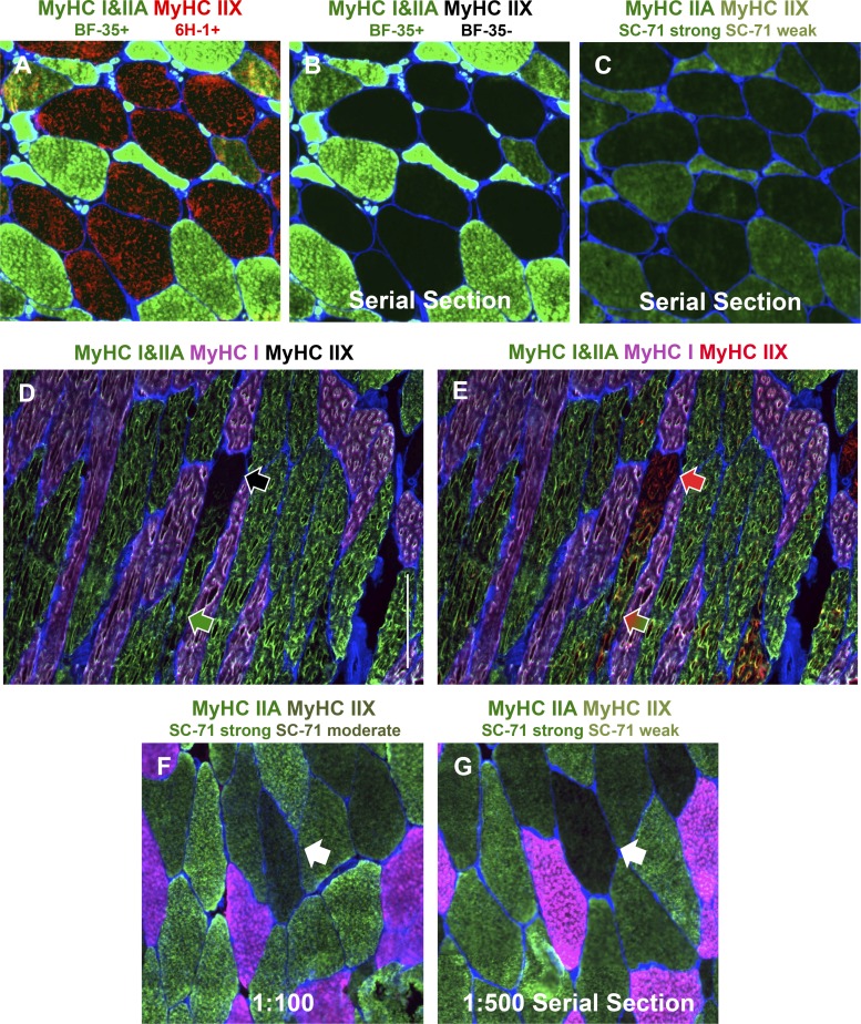
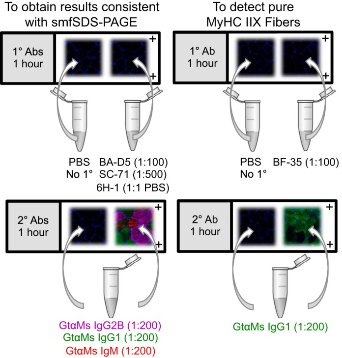
Skeletal muscle myosin heavy chain (MyHC) fiber type composition is a critical determinant of overall muscle function and health. Various approaches interrogate fiber type at the single cell, but the two most commonly utilized are single-muscle fiber sodium dodecyl sulfate-polyacrylamide gel electrophoresis (smfSDS-PAGE) and fluorescent immunohistochemistry (IHC). Although smfSDS-PAGE is generally considered the "gold standard," IHC is more commonly used because of its time-effectiveness and relative ease. Unfortunately, there is lingering inconsistency on how best to accurately and quickly determine fiber type via IHC and an overall misunderstanding regarding pure fiber type proportions, specifically the abundance of fibers exclusively expressing highly glycolytic MyHC IIX in humans. We therefore 1) present information and data showing the low abundance of pure MyHC IIX muscle fibers in healthy human skeletal muscle and 2) leverage this information to provide straightforward protocols that are informed by human biology and employ inexpensive, easily attainable antibodies for the accurate determination of fiber type.

Keywords: MyHC; SDS-PAGE; immunohistochemistry.


Conflict of interest statement

No conflicts of interest, financial or otherwise, are declared by the authors.

Figures

Fig. 1.
Fig. 1.
Fluorescent immunohistochemistry (IHC; A and B) and single-muscle fiber sodium dodecyl sulfate-polyacrylamide gel electrophoresis (smfSDS-PAGE; C and D) for myosin heavy chain (MyHC) fiber typing in vastus lateralis biopsy samples from a cohort of healthy adult men and women (n = 22; 10 men/12 women). A: representative IHC image of MyHC I (green), MyHC IIA (red), and MyHC IIA/IIX (blue/red) fibers. Scale bar, 50 µm. B: quantification of muscle fiber type via IHC (2,552 total fibers), with gray box emphasizing low MyHC IIX abundance. C: representative image of a smfSDS-PAGE gel showing the continuum of MyHC fiber types. D: quantification of smfSDS-PAGE data from mechanically dissected single muscle fibers (876 total fibers) from the same 22 subjects analyzed in B, with gray box emphasizing low MyHC IIX abundance. Skeletal muscle biopsy samples were obtained under resting conditions, and smfSDS-PAGE was conducted as described by our laboratories in detail elsewhere (36). IHC was conducted in the Toth laboratory according to the methods described in the text, and images were captured at ×20 magnification. Std, standard.
Fig. 2.
Fig. 2.
Immunohistochemistry (IHC) demonstrating specificity of myosin heavy chain (MyHC) antibodies and regional MyHC accumulation. A–C: representative images of sections from a muscle biopsy from a spinal cord-injured man (50 yr old, American Spinal Cord Injury Association classification “A”, 13 yr postinjury), taken at the same exposure. A and B: fiber typing using BF-35 (MyHC I and IIA, green) and 6H-1 (MyHC IIX, red) (A) compared with BF-35 only (B) on the same section. C: a serial section of the same region as in A and B showing fibers strongly (MyHC IIA) and weakly (MyHC IIX) positive for SC-71 (green), demonstrating the preferential affinity of the SC-71 antibody for MyHC IIA but weak affinity for MyHC IIX. D and E: representative images of a longitudinal muscle section from a young healthy man showing a muscle fiber that transitions from pure MyHC IIX [negative for BF-35, no fluorescence (black arrow in D), positive for 6H-1 (red arrow in E)] to MyHC IIA/IIX [positive for BF-35 (green arrow in D) and 6H-1 (green and red arrow in E)]. Scale bar, 300 µm. F and G: SC-71 applied to a muscle sample from a healthy young subject at 1:100 (F) and 1:500 (G) dilutions, illustrating the effect of antibody concentration on cross-reactivity (white arrows point to differential reactivity in the same fiber on serial sections). MyHC I is pink in D–G. Muscle tissue was obtained from the Bamman laboratory (A–C) and the University of Kentucky Center for Muscle Biology Tissue Bank (https://www.uky.edu/chs/muscle/bank), and IHC was conducted in the Peterson laboratory (D and E). Fiber borders (laminin) are in blue, and all images were captured at ×10 magnification.
Fig. 3.
Fig. 3.
Visual representations of recommended immunohistochemistry (IHC) fiber-typing protocols. Muscle fiber borders (laminin, blue) are shown on all images for illustrative purposes. Left: pink is myosin heavy chain (MyHC) I, green is MyHC IIA, and red is MyHC IIX. Right: green is MyHC I and/or IIA, and no fluorescence is MyHC IIX.

References

    1. Andersen JL, Klitgaard H, Bangsbo J, Saltin B. Myosin heavy chain isoforms in single fibres from m. vastus lateralis of soccer players: effects of strength-training. Acta Physiol Scand 150: 21–26, 1994. doi:10.1111/j.1748-1716.1994.tb09655.x. - DOI - PubMed
    1. Andersen JL, Klitgaard H, Saltin B. Myosin heavy chain isoforms in single fibres from m. vastus lateralis of sprinters: influence of training. Acta Physiol Scand 151: 135–142, 1994. doi:10.1111/j.1748-1716.1994.tb09730.x. - DOI - PubMed
    1. Andersen JL, Mohr T, Biering-Sørensen F, Galbo H, Kjaer M. Myosin heavy chain isoform transformation in single fibres from m. vastus lateralis in spinal cord injured individuals: effects of long-term functional electrical stimulation (FES). Pflugers Arch 431: 513–518, 1996. doi:10.1007/BF02191897. - DOI - PubMed
    1. Andersen JL, Terzis G, Kryger A. Increase in the degree of coexpression of myosin heavy chain isoforms in skeletal muscle fibers of the very old. Muscle Nerve 22: 449–454, 1999. doi:10.1002/(SICI)1097-4598(199904)22:4<449:AID-MUS4>3.0.CO;2-2. - DOI - PubMed
    1. Arevalo JA, Lynn SK, Bagley JR, Brown LE, Costa PB, Galpin AJ. Lower-limb dominance, performance, and fiber type in resistance-trained men. Med Sci Sports Exerc 50: 1054–1060, 2018. doi:10.1249/MSS.0000000000001533. - DOI - PubMed

Publication types

MeSH terms

Substances

LinkOut - more resources