. 2020 Jan;577(7789):275-279.
doi: 10.1038/s41586-019-1759-1. Epub 2019 Nov 7.

Structure of the human metapneumovirus polymerase phosphoprotein complex

Affiliations

Structure of the human metapneumovirus polymerase phosphoprotein complex

Junhua Pan et al. Nature. 2020 Jan.

Abstract

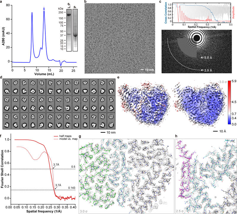
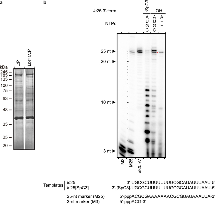
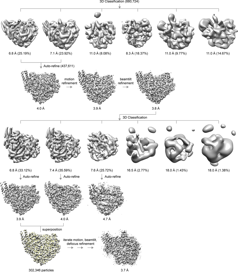
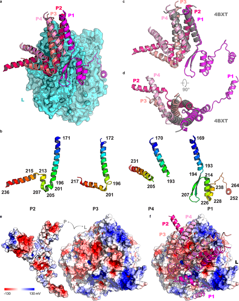
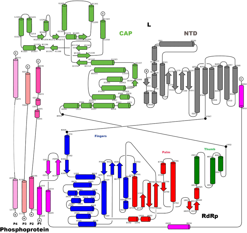
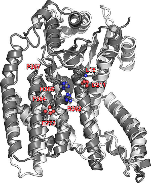
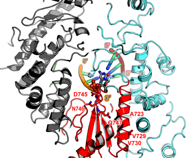
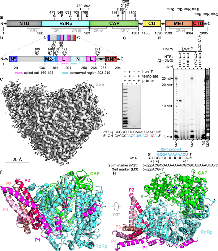
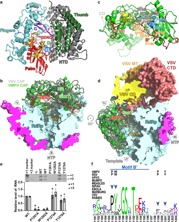
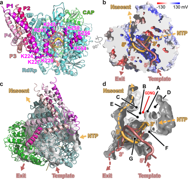
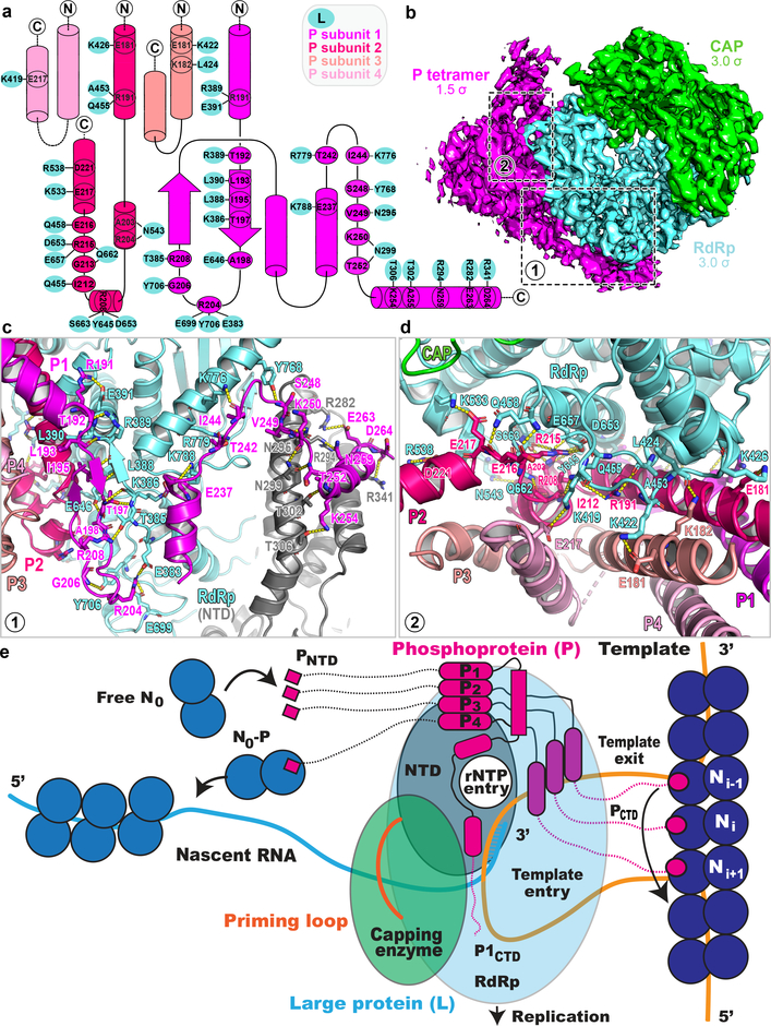
Respiratory syncytial virus (RSV) and human metapneumovirus (HMPV) cause severe respiratory diseases in infants and elderly adults1. No vaccine or effective antiviral therapy currently exists to control RSV or HMPV infections. During viral genome replication and transcription, the tetrameric phosphoprotein P serves as a crucial adaptor between the ribonucleoprotein template and the L protein, which has RNA-dependent RNA polymerase (RdRp), GDP polyribonucleotidyltransferase and cap-specific methyltransferase activities2,3. How P interacts with L and mediates the association with the free form of N and with the ribonucleoprotein is not clear for HMPV or other major human pathogens, including the viruses that cause measles, Ebola and rabies. Here we report a cryo-electron microscopy reconstruction that shows the ring-shaped structure of the polymerase and capping domains of HMPV-L bound to a tetramer of P. The connector and methyltransferase domains of L are mobile with respect to the core. The putative priming loop that is important for the initiation of RNA synthesis is fully retracted, which leaves space in the active-site cavity for RNA elongation. P interacts extensively with the N-terminal region of L, burying more than 4,016 Å2 of the molecular surface area in the interface. Two of the four helices that form the coiled-coil tetramerization domain of P, and long C-terminal extensions projecting from these two helices, wrap around the L protein in a manner similar to tentacles. The structural versatility of the four P protomers-which are largely disordered in their free state-demonstrates an example of a 'folding-upon-partner-binding' mechanism for carrying out P adaptor functions. The structure shows that P has the potential to modulate multiple functions of L and these results should accelerate the design of specific antiviral drugs.


Conflict of interest statement

Competing interests. The authors declare no competing interests

Figures

Extended Data Fig. 1 |
Extended Data Fig. 1 |. Purification of HMPV L:P and structure determination using cryo-EM
a, Representative size exclusion chromatogram of the L:P complex (these experiments were repeated more than 5 times). Fractions indicated by an arrow were collected and concentrated to 0.85 mg/mL and used for cryo EM analysis. Inset: SDS PAGE followed by Coomassie blue staining of the purified samples. Also shown: free P protein separated from L:P complex by heparin chromatography (For gel source data, see Supplementary Fig. 1). b, Raw micrograph of HMPV-L:P particles recorded in vitreous ice. Scale bar 10 nm. c, Power spectrum of the image shown in panel (b). We limited the high resolution for fitting to a spatial frequency of 1/5.0 Å and 1/2.9 Å marks the highest spacing to which CTF rings were successfully fit. d, 2D classes and “self-consistency check” for the cryo-EM 3D reconstruction. In each box over the three rows, the upper panel shows one 2D class average, whilst the lower panel shows the corresponding projection from the initial 3D model. e, Local resolution of the cryo-EM density map. Variations in local resolution are color- coded from blue (3.0 Å) to red (5.9 Å), computed with Resmap. f, Fourier Shell Correlation (FSC) of the cryo-EM map as a function of the spatial frequency. The gold standard resolution is 3.7 Å based on the FSC=0.143 criterion, consistent with the model to map correlation (0.5 criterion). g, Example of the electron density map that allowed model building. The region shown is at an interface between the RdRp and capping domain. The map is shown as a gray mesh, contoured at a level of 3 σ. The atomic model is shown as sticks with residues from RdRp colored in cyan (NTD in grey) and in green for the capping domain. h, The region shown is the three-stranded β-sheet at the interface between the RdRp (cyan sticks) and the phosphoprotein (magenta sticks). The map is shown as a gray mesh, contoured at a level of 2.5 σ. We observed a nearly identical structure of the L:P complex in a reconstruction obtained by premixing the L:P complex with fully phosphorylated P, indicating that potential exchange of P affected neither the formation nor the structure of the L:P complex.
Extended Data Fig. 2 |
Extended Data Fig. 2 |. RdRp activity assay.
a, SDS PAGE of HMPV wild type L:P, LD745A:P purified for RdRp activity assays. Proteins were purified by metal affinity, TEV cleavage of Histidine-tag followed by reverse His-tag affinity purification and size exclusion chromatography. b, Analysis of the 3′ extension activity of HMPV polymerase using the le25 RNA template. Reactions were performed with rNTPs (0.5 mM each of rUTP, rGTP and rCTP), 20 μM rATP and 20 nM [α-32P] rATP. When a 3’-modified le25 (le25[SpC3], three-carbon spacer group linked to the 3’ extremity) was used as a template, synthesis of products greater than 25 nt was greatly reduced compared to le25. When only [α-32P]rATP and no other rNTP was supplied, only a product with size greater than 25 nt was observed. This result shows that the L:P complex was capable of modifying the 3’ terminus of the template, in addition to engaging in de novo initiation at the promoter. The radiolabeled RNA products were visualized by phosphorimaging. Data are representative of three independent experiments. For gel source data, see Supplementary Fig. 1.
Extended Data Fig. 3 |
Extended Data Fig. 3 |. Flow-chart depicting structure determination using cryo-EM.
Please see methods sections for details.
Extended Data Fig. 4 |
Extended Data Fig. 4 |. Phosphoprotein tetramer in complex with L.
a, The L protein (cyan) is represented as a molecular surface and the tetrameric P protein subunits are represented as ribbons, following the color codes in Figs. 1–3 (P1 in magenta, P2 in hot pink, P3 in salmon and P4 in pink). b, Structures adopted by the four individual P subunits bound to L, colored as a blue to red “rainbow” from the N- to the C- terminal ends. Secondary structures boundaries are noted for each subunit. c and d, Superposition of the tetramerization helices in the context of the L:P complex and the free P protein. Structures are represented as colored ribbons with the free phosphoprotein coiled-coil (PDB access code 4BXT) colored in gray and the four P subunits reported in this work colored according to Fig. 1 (P1 in magenta; P2, hot pink; P3, salmon and P4, pink). The r.m.s.d. of the superimposition is 1.13 Å over 88 α-carbon atoms. e, View of the complex where L and P have been pulled apart to display electrostatic surfaces. f, Overall view of the L:P complex with P shown as ribbons and L as electrostatic surface. The P tetramer consists of subunits P1(magenta), P2 (hotpink), P3 (salmon), and P4 (pink).
Extended Data Figure 5 |
Extended Data Figure 5 |. Topology of the L:P complex.
Topological depiction of the secondary structure elements of L and P. Helices are depicted as tubes and strands as arrows. The color code is the same as in Fig. 1. The RdRp domain and its subdomains and the capping domain are colored as in Figs. 1 and 2: NTD in grey, finger in blue, palm in red, thumb in dark green and CAP in green. The four subunits of the phosphoprotein P1, P2, P3 and P4 are colored as in Fig. 1. Secondary structure boundaries are indicated.
Extended Data Fig. 6 |
Extended Data Fig. 6 |. View of the N-terminal domain (NTD).
NTD is displayed as grey ribbons (following Fig. 2a colors), with evolutionary conserved residues clustered near the rNTP entry tunnel playing a role in transcription, represented as sticks and labeled. Colored in lighter grey is the equivalent region of the VSV_L superimposed to HMPV_L.
Extended Data Fig. 7 |
Extended Data Fig. 7 |. Model for an elongation complex stalled by the addition of ALS-8176 5’-triphosphate.
ALS-8176 5’-triphosphate is a nucleoside triphosphate analog against RSV and HMPV currently in phase 2 clinical trials. The 744GDNQ catalytic motif and positions (A789V, L795I and I796V) of which mutations conferred resistance (identified by passaging RSV) are mapped onto the HMPV-L structure (respectively corresponding to A723, V729 and V730) and displayed as sticks. These conservative mutations probably affect inhibitor binding by inducing a slight repositioning of the helix, due to altered hydrophobic contacts with neighboring helices. Please refer also to the sequence alignment displayed in Supplementary Fig. 2. Protein is colored according to Fig. 2a, and the template and nascent RNA strands according to Fig. 3a.
Extended Data Fig. 8 |
Extended Data Fig. 8 |. MS2 spectrum of the Ser148 P phosphopeptide.
(a) One MS2 spectrum used for identification of the phosphorylated P peptide 142DALDLLS#DNEEEDAESSILTFEER is displayed. Tandem mass spectrum (top) and deviation (bottom) allowed detection of phosphorylation (symbol #) at site Ser148. Peptides fragmented from the N-terminus (b-fragments) and C-terminus (y-fragments) are colored in blue and red, respectively. (b) y and b ion series m/z identified in the spectrum (a) and their deviation from theoretical m/z are displayed in the Table. The present pattern of phosphorylation agrees with observations showing that phosphorylation of the peptide comprising residues 100–120 (ref 44) of RSV-P - in particular phosphorylation of Thr108 (ref 45) corresponding to Ser148 of HMPV-P (Extended Data Fig. 9)) - controls its interaction with the M2–1 protein.
Extended Data Fig. 9 |
Extended Data Fig. 9 |. Structure-based sequence alignment of the phosphoprotein from HMPV (labeled HMPV-A, strain CAN97-83) and other known pneumoviral P proteins:
HMPV-B, human metapneumovirus subgroup B; HRSV-A and B, human respiratory syncytial virus subgroup A and B respectively; BRSV, bovine respiratory syncytial virus; PVM, pneumonia virus of mice; AMPV-A and AMPV-C, avian metapneumovirus subgroup A and C respectively. Sequences accession codes for the alignment HMPV-A: AAQ67693.1 (used in this work), HMPV-B: AAQ67684.1, HRSV-A: AAX23990.1, HRSV-B: AAR14262.1, BRSV: AAL49395.1, PVM: AAW79177.1, AMPV-A: AAT68644.1 and AMPV-C: AAT86110.1. The secondary structure of HMPV_P subunit P1 (this work) is displayed above the alignment. Phosphorylation sites are highlighted in brown. Positively-charged residues of HMPV_P are shaded in blue, negatively charged residues in magenta and hydrophobic residues 29 to 135 in yellow. The conserved region containing hydrophobic residues critical for L:P interactions are highlighted in green. Structural alignment of P from HMPV and RSV showed similar overall tetramer organization. However, differences are observed in subunit P1 with an r.m.s.d. of 2.24 Å over 82 residues. Although P is in general more mobile with weaker densities and higher B factors compared to L, the region following the beta-hairpin (residues 175–215 in HMPV) does adopt a slightly different conformation compared to RSV P1. Subunit P3 has an r.m.s.d. of 1.94 Å over 45 residues due to a slightly tilted C-terminal helix compared to RSV. Subunit P2 is most similar with an r.m.s.d. of 0.92 Å over 56 residues. Subunit P4 has an r.m.s.d. of 1.33 Å over 47 residues. The eight residues of HRSV-P, that are crucial for interacting with HRSV-L and whose substitutions impair viral replication, are shaded in dark green (data from reference 16). With the exception of Asn189 (HRSV-P) where a deletion is present in HMPV-P, these residues are conserved in HMPV-P and other known pneumoviral P proteins.
Figure 1.
Figure 1.. Overall structure of the HMPV L:P complex.
Domain organization of L (a) and P (b) outlining conserved regions and motifs. Regions of HMPV-P predicted to interact with N0, M2–1, L and RNP, and phosphorylation sites are indicated. (c) Primer elongation assay. Sequences of the 18 nt RNA template and for the 5 nt primer are shown with nascent RNA in blue and radiolabel incorporation sites in red. This experiment was performed a total of four times with two different buffer conditions. (d) RdRp activity assay using the “le14” RNA template. Sequences for the 5’-triphosphorylated 25 nt and 3 nt markers and the 12 nt product are indicated. The 3’dGTP chain terminator is labeled “G”. Radiolabeled UMP is in red. Data are representative of three independent experiments. For gel source data, see Supplementary Fig. 1. (e) Overview of the HMPV-L:P cryo-EM 3D reconstruction (f) Overview of the L:P atomic model. RdRp: cyan, Capping domain: green, phosphoprotein tetramer subunits P1 magenta, P2 hot pink, P3 salmon and P4 pink. Atoms from the “GDNQ” motif in the RdRp and from the “HR” motif in the capping domains are shown as colored spheres (g) Rotated view of the L:P atomic model.
Figure 2.
Figure 2.. Structure of the HMPV L protein.
(a) The RdRp viewed in its “front” orientation. Fingers subdomain: blue; palm: red and thumb: green, NTD: grey. RdRp motifs A-G are shown. (b) The capping domain of L (green ribbons) of HMPV-L. A superposition of HMPV-L with VSV-L3 (grey ribbons) highlights the difference between conformations of the putative priming loops: putative priming loop of HMPV-L (including Thr1192 of the GxxT motif): red and of VSV-L (1157–1173): gold. P: magenta. (c) RNA capping motifs A’-E’. (d) Overall view of HMPV-L with VSV-L superimposed, highlighting the mobile appendage (displayed in the VSV-L orientation, but disordered in the present structure) and rNTP (S) entry, template (T) and nascent (U) RNA tunnels. (e) Impact of substitutions in aromatic and proline residues in the putative priming loop of RSV-L on RNA production from the +1 and +3 sites. The mean and standard error of three independent experiments are indicated. (f) Sequence conservation in the putative priming loop of L. The four residues that were mutated (panel e) are indicated by arrows. A proline residue (P1186 in HMPV-L) important for an early stage of RNA synthesis is conserved in Pneumoviridae, Paramyxoviridae and Filoviridae but not in Rhabdoviridae.
Figure 3.
Figure 3.. Model for RNA elongation by L:P from Pneumoviridae.
(a) Model for HMPV L:P elongation complex. Template strand: firebrick, nascent strand: orange. Residues from L (Lys307, Arg313, Arg788) and from the C-terminus of subunit P1 (Lys at positions 224, 227, 229, 243, 250, 254 and 256 and Arg241) form a positively-charged arch attracting rNTPs to the rNTP entry tunnel. (b) Cut-out view of L showing electrostatic surfaces (blue: positive and red: negative) and paths followed by the template and nascent RNA strands. (c) Tunnels and interior cavities of L depicted as a white surface enclosed by the L:P complex (ribbons). Cut open are various tunnel openings leading to the exterior. (d) Magnified view of the tunnel that traverses the L protein. Functional motifs A-G and catalytic residues 744GDNQ are indicated.
Figure 4.
Figure 4.. The P homo-tetramer and L:P interactions.
(a) Interactions between residues from P1 (magenta), P2 (hotpink), P3 (salmon), P4 (pink) of P and the RdRp (cyan). (b) The electron density map (RdRp: cyan, capping domain: green and P: magenta). (c) A β-hairpin of P1 forms an antiparallel β-sheet with residues 383–391 of the fingers subdomains of L (cyan). Polar contacts between P1 and L are indicated by dashes. (d) interactions between the coiled-coil of P and other parts of P2, P3 and P4 with L. (e) Model for RNA replication by L:P. Genomic viral RNA, extruded from three adjacent N units from the RNP, inserts as a loop of ~27 nts into the RNA entry tunnel and back through the template exit tunnel. Genomic RNA displacement is achieved by three PCTD of phosphoprotein subunits P2, P3 and P4 (while P1 plays a structural role). Concomitantly, four PNTD helices, flexibly linked to the tetramerization domain, bring four new free N0 subunits towards the nascent RNA exit tunnel for encapsidation.

Similar articles

Cited by

References

    1. van den Hoogen BG, de Jong JC, Groen J, Kuiken T, de Groot R, Fouchier RA, Osterhaus AD A newly discovered human pneumovirus isolated from young children with respiratory tract disease. Nat. Med 7,719–24 (2001). - PMC - PubMed
    1. Neubauer J, Ogino M, Green TJ, Ogino T Signature motifs of GDP polyribonucleotidyltransferase, a non-segmented negative strand RNA viral mRNA capping enzyme domain in the L protein are required for covalent enzyme-pRNA intermediate formation. Nucleic Acids Res. 44(1) 330–341 (2016). - PMC - PubMed
    1. Liang B, Li Z, Jenni S, Rahmeh AA, Morin BM, Grant T, Grigorieff N, Harrison SC, Whelan SP Structure of the L Protein of Vesicular Stomatitis Virus from Electron Cryomicroscopy. Cell 162, 314–327. doi: 10.1016/j.cell.2015.06.018 (2015). - DOI - PMC - PubMed
    1. Pflug A, Guilligay D, Reich S, Cusack S Structure of Influenza A polymerase bound to the viral RNA promoter. Nature 516, 355–60 (2014). - PubMed
    1. Paesen GC, Collet A, Sallamand C, Debart F, Vasseur JJ, Canard B, Decroly E, Grimes JM X-ray structure and activities of an essential Mononegavirales L-protein domain. Nat. Commun 6, 8749. doi: 10.1038/ncomms9749 (2015). - DOI - PMC - PubMed

Publication types

Substances