

Global Burden of Small Vessel Disease-Related Brain Changes on MRI Predicts Cognitive and Functional Decline
- PMID: 31699021
- PMCID: PMC6924941
- DOI: 10.1161/STROKEAHA.119.026170
Global Burden of Small Vessel Disease-Related Brain Changes on MRI Predicts Cognitive and Functional Decline
Abstract
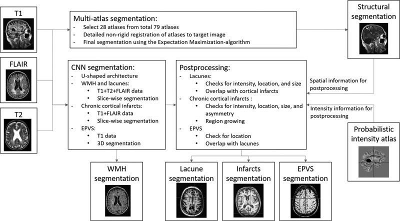
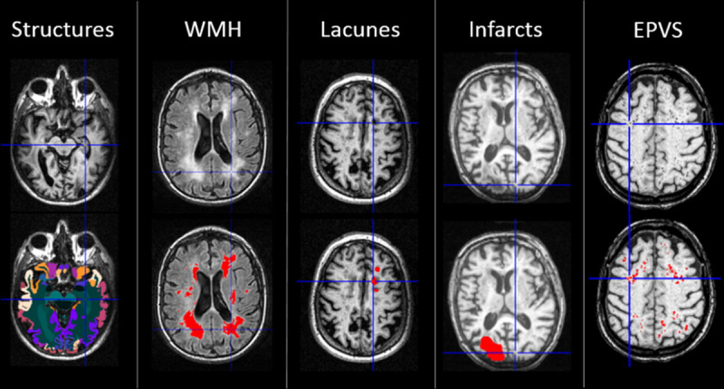
Background and Purpose- Cerebral small vessel disease is characterized by a wide range of focal and global brain changes. We used a magnetic resonance imaging segmentation tool to quantify multiple types of small vessel disease-related brain changes and examined their individual and combined predictive value on cognitive and functional abilities. Methods- Magnetic resonance imaging scans of 560 older individuals from LADIS (Leukoaraiosis and Disability Study) were analyzed using automated atlas- and convolutional neural network-based segmentation methods yielding volumetric measures of white matter hyperintensities, lacunes, enlarged perivascular spaces, chronic cortical infarcts, and global and regional brain atrophy. The subjects were followed up with annual neuropsychological examinations for 3 years and evaluation of instrumental activities of daily living for 7 years. Results- The strongest predictors of cognitive performance and functional outcome over time were the total volumes of white matter hyperintensities, gray matter, and hippocampi (P<0.001 for global cognitive function, processing speed, executive functions, and memory and P<0.001 for poor functional outcome). Volumes of lacunes, enlarged perivascular spaces, and cortical infarcts were significantly associated with part of the outcome measures, but their contribution was weaker. In a multivariable linear mixed model, volumes of white matter hyperintensities, lacunes, gray matter, and hippocampi remained as independent predictors of cognitive impairment. A combined measure of these markers based on Z scores strongly predicted cognitive and functional outcomes (P<0.001) even above the contribution of the individual brain changes. Conclusions- Global burden of small vessel disease-related brain changes as quantified by an image segmentation tool is a powerful predictor of long-term cognitive decline and functional disability. A combined measure of white matter hyperintensities, lacunar, gray matter, and hippocampal volumes could be used as an imaging marker associated with vascular cognitive impairment.
Keywords: brain; cerebral small vessel diseases; humans; image processing, computer assisted; neuropsychology.
Figures
Comment in
-
Global small vessel disease brain changes predict cognitive decline.Nat Rev Neurol. 2020 Jan;16(1):2-3. doi: 10.1038/s41582-019-0296-8. Nat Rev Neurol. 2020. PMID: 31776476 No abstract available.
References
-
- Wardlaw JM, Smith EE, Biessels GJ, Cordonnier C, Fazekas F, Frayne R, et al. Standards for Reporting Vascular Changes on Neuroimaging (STRIVE v1) Neuroimaging standards for research into small vessel disease and its contribution to ageing and neurodegeneration. Lancet Neurol. 2013;12:822–838. doi: 10.1016/S1474-4422(13)70124-8. - PMC - PubMed
-
- Arba F, Quinn TJ, Hankey GJ, Lees KR, Wardlaw JM, Ali M, et al. VISTA Collaboration. Enlarged perivascular spaces and cognitive impairment after stroke and transient ischemic attack. Int J Stroke. 2018;13:47–56. doi: 10.1177/1747493016666091. - PubMed
-
- Jokinen H, Gouw AA, Madureira S, Ylikoski R, van Straaten EC, van der Flier WM, et al. LADIS Study Group. Incident lacunes influence cognitive decline: the LADIS study. Neurology. 2011;76:1872–1878. doi: 10.1212/WNL.0b013e31821d752f. - PubMed
-
- Jokinen H, Lipsanen J, Schmidt R, Fazekas F, Gouw AA, van der Flier WM, et al. LADIS Study Group. Brain atrophy accelerates cognitive decline in cerebral small vessel disease: the LADIS study. Neurology. 2012;78:1785–1792. doi: 10.1212/WNL.0b013e3182583070. - PubMed
