Review
. 2019 Dec;19(12):e405-e421.
doi: 10.1016/S1473-3099(19)30312-3. Epub 2019 Nov 5.

Global guideline for the diagnosis and management of mucormycosis: an initiative of the European Confederation of Medical Mycology in cooperation with the Mycoses Study Group Education and Research Consortium

Oliver A Cornely  1 Ana Alastruey-Izquierdo  2 Dorothee Arenz  3 Sharon C A Chen  4 Eric Dannaoui  5 Bruno Hochhegger  6 Martin Hoenigl  7 Henrik E Jensen  8 Katrien Lagrou  9 Russell E Lewis  10 Sibylle C Mellinghoff  3 Mervyn Mer  11 Zoi D Pana  12 Danila Seidel  3 Donald C Sheppard  13 Roger Wahba  14 Murat Akova  15 Alexandre Alanio  16 Abdullah M S Al-Hatmi  17 Sevtap Arikan-Akdagli  18 Hamid Badali  19 Ronen Ben-Ami  20 Alexandro Bonifaz  21 Stéphane Bretagne  16 Elio Castagnola  22 Methee Chayakulkeeree  23 Arnaldo L Colombo  24 Dora E Corzo-León  25 Lubos Drgona  26 Andreas H Groll  27 Jesus Guinea  28 Claus-Peter Heussel  29 Ashraf S Ibrahim  30 Souha S Kanj  31 Nikolay Klimko  32 Michaela Lackner  33 Frederic Lamoth  34 Fanny Lanternier  35 Cornelia Lass-Floerl  33 Dong-Gun Lee  36 Thomas Lehrnbecher  37 Badre E Lmimouni  38 Mihai Mares  39 Georg Maschmeyer  40 Jacques F Meis  41 Joseph Meletiadis  42 C Orla Morrissey  43 Marcio Nucci  44 Rita Oladele  45 Livio Pagano  46 Alessandro Pasqualotto  47 Atul Patel  48 Zdenek Racil  49 Malcolm Richardson  50 Emmanuel Roilides  12 Markus Ruhnke  51 Seyedmojtaba Seyedmousavi  52 Neeraj Sidharthan  53 Nina Singh  54 János Sinko  55 Anna Skiada  56 Monica Slavin  57 Rajeev Soman  58 Brad Spellberg  59 William Steinbach  60 Ban Hock Tan  61 Andrew J Ullmann  62 Jörg J Vehreschild  63 Maria J G T Vehreschild  64 Thomas J Walsh  65 P Lewis White  66 Nathan P Wiederhold  67 Theoklis Zaoutis  68 Arunaloke Chakrabarti  69 Mucormycosis ECMM MSG Global Guideline Writing Group
Affiliations
Review

Global guideline for the diagnosis and management of mucormycosis: an initiative of the European Confederation of Medical Mycology in cooperation with the Mycoses Study Group Education and Research Consortium

Oliver A Cornely et al. Lancet Infect Dis. 2019 Dec.

Abstract

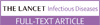
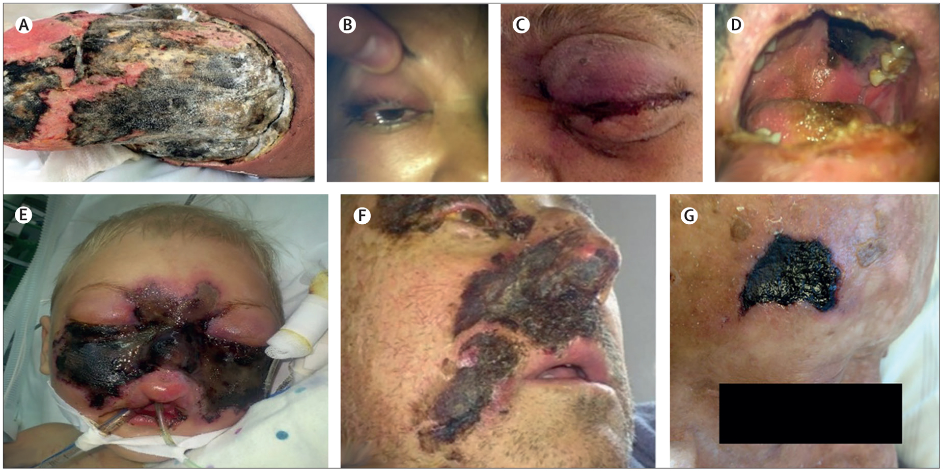
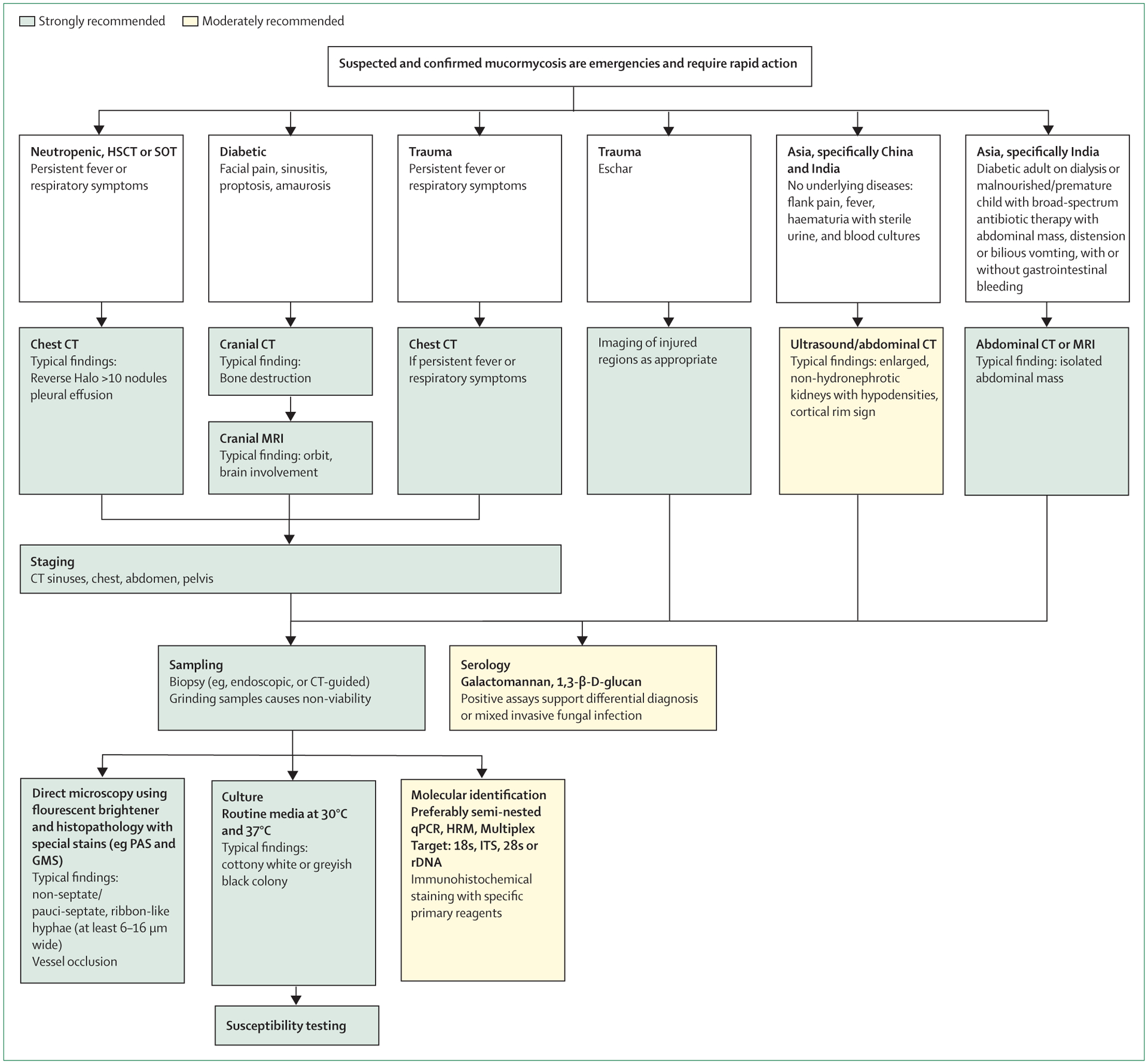
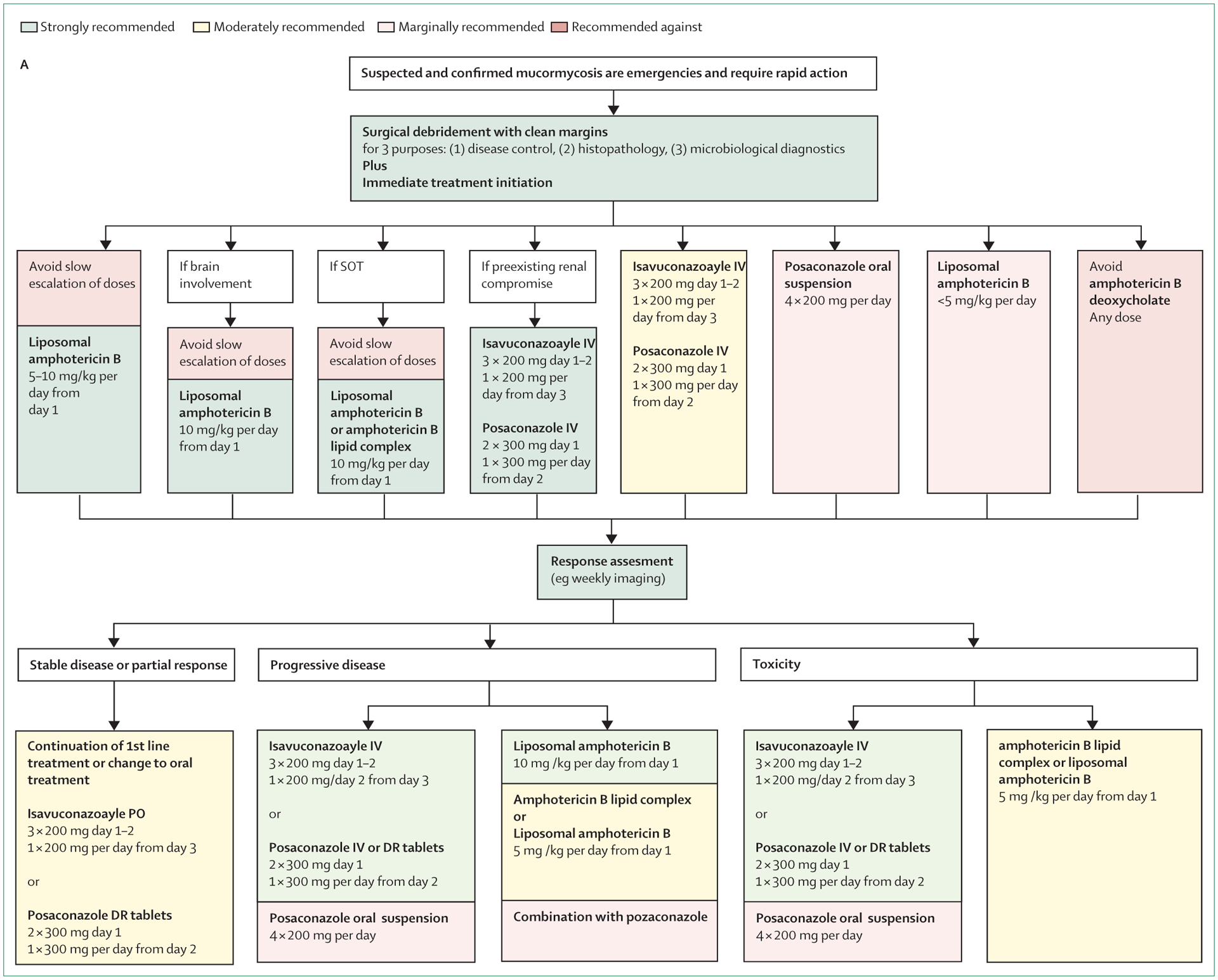
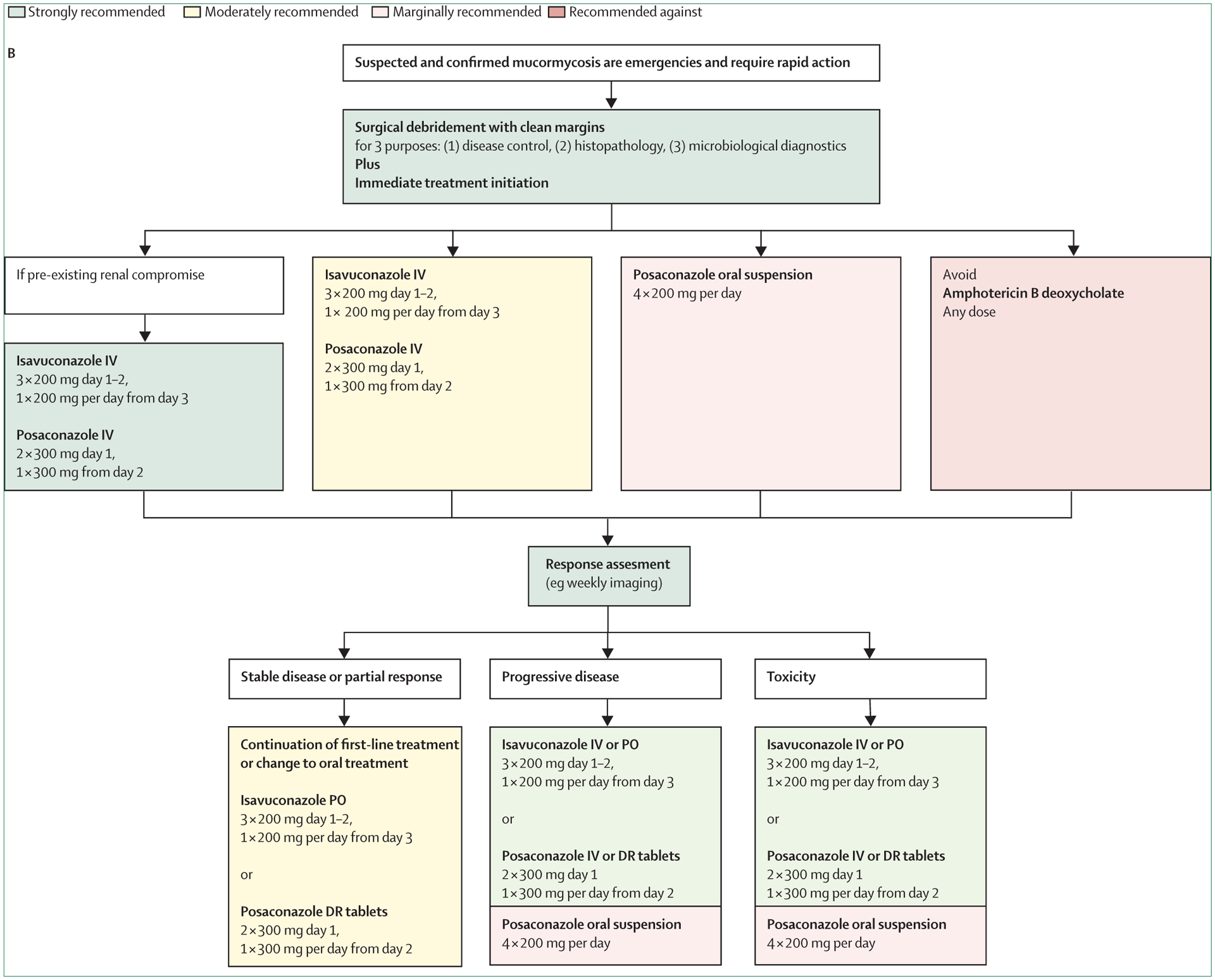
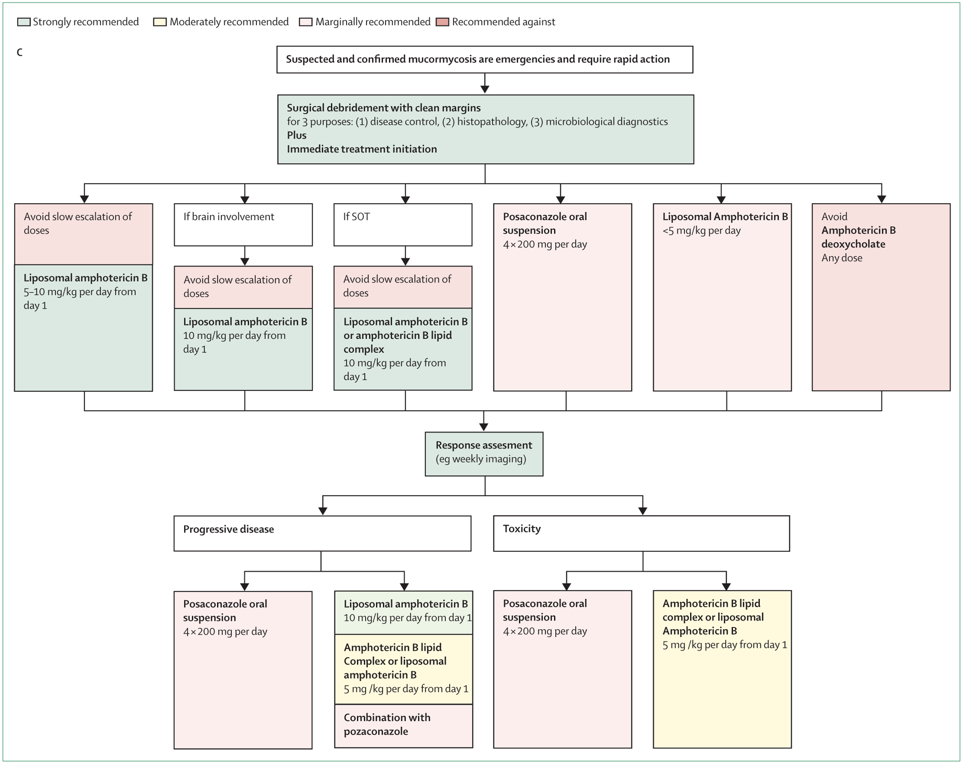
Mucormycosis is a difficult to diagnose rare disease with high morbidity and mortality. Diagnosis is often delayed, and disease tends to progress rapidly. Urgent surgical and medical intervention is lifesaving. Guidance on the complex multidisciplinary management has potential to improve prognosis, but approaches differ between health-care settings. From January, 2018, authors from 33 countries in all United Nations regions analysed the published evidence on mucormycosis management and provided consensus recommendations addressing differences between the regions of the world as part of the "One World One Guideline" initiative of the European Confederation of Medical Mycology (ECMM). Diagnostic management does not differ greatly between world regions. Upon suspicion of mucormycosis appropriate imaging is strongly recommended to document extent of disease and is followed by strongly recommended surgical intervention. First-line treatment with high-dose liposomal amphotericin B is strongly recommended, while intravenous isavuconazole and intravenous or delayed release tablet posaconazole are recommended with moderate strength. Both triazoles are strongly recommended salvage treatments. Amphotericin B deoxycholate is recommended against, because of substantial toxicity, but may be the only option in resource limited settings. Management of mucormycosis depends on recognising disease patterns and on early diagnosis. Limited availability of contemporary treatments burdens patients in low and middle income settings. Areas of uncertainty were identified and future research directions specified.


Figures

Figure 1:
Figure 1:. Cutaneous and rhino-orbito-cerebral mucormycosis
(A) Extensive primary cutaneous mucormycosis of the left leg due to Apophysomyces variabilis, after a car accident. (B) Erythematous skin, ptosis, palpebral oedema, limited ocular motility, and right maxillary pain, 6 days after symptom onset in uncontrolled diabetes. (C) Proptosis, palpebral erythema, and cavernous sinus syndrome, 7 days after symptom onset in uncontrolled diabetes. (D) Necrotic, purulent palatal ulcer and cavernous sinus syndrome, 8 days after symptom onset in uncontrolled diabetes. (E) Rhinocerebral mucormycosis in a female child, 2 years old with acute lymphoblastic leukaemia and lethal outcome. (F) 52-year-old man with persistent neutropenia post chemotherapy, sinusitis, and skin necrosis. (G) Black eschar as typical skin lesion in mucormycosis; one of several lesions on the right forehead, ear and cheek in a non-diabetic, haematopoietic stem cell transplant recipient with pansinusitis due to Lichtheimia corymbifera. Image A courtesy of Alexandro Bonifaz, images B–D courtesy of Dora E Corzo-León, images E and F courtesy of Valentina Arsic Arsenijevic, Belgrade, Serbia, and image G courtesy of University Hospital Cologne. We obtained written permission from patients or parents respectively to publish images, and from ethics committee as appropriate per local regulation.
Figure 2:
Figure 2:. Diagnostic pathway for mucormycosis
Depending on the geographical location not all recommended tests might have regulatory approval for use in clinical settings. HSCT=haematopoietic stem cell translplantation. SOT=solid organ transplantation. PAS=periodic acid Schiff. GMS=Grocott-Gomori’s methenamine-silver strain. qPCR=quantitative PCR. HRM=high resolution melting. ITS=internal transcribed spacer. rDNA=ribosomal DNA.
Figure 3:
Figure 3:. Radiographic signs of mucormycosis
Four imaging signs can suggest pulmonary mucormycosis in an appropriate clinical setting. (A) Halo sign on CT, a ring of ground glass opacity surrounding a nodular infiltrate, which pathophysiologically represents a region of ischaemia, and which is also typical of invasive pulmonary aspergillosis (arrow). (D & B) Reversed halo sign on CT, also known as inversed halo or atoll sign, an area of ground glass opacity surrounded by a ring of consolidation (arrow). (E) Hypodense sign on MRI, T1 weighted, a central hypodensity in a lung consolidation or nodule, corresponding to a central area of necrosis caused by vascular obstruction with secondary lung infarction and sequestration. Magnetic resonance imaging shows pulmonary nodule with central hypodensity in right upper lobe (arrow), corresponding to a central area of necrosis caused by vascular obstruction with secondary lung infarct and sequestration. (C) Vascular occlusion sign on CT angiography, defined as interrupted vessel at the border of a focal lesion without depiction of the vessel inside the lesion or peripheral to the lesion (arrow). Particularly aggressive forms of mucormycosis are F. Contiguous spread on CT, presence of a mass or consolidation exhibiting invasion of adjacent organs by traversing tissue planes, including the diaphragm, chest wall, pleura, and spleen. (G) Typical rapidly progressive pulmonary mucormycosis on CT, associated with clinical deterioration. Day 8 and Day 15 CT scans showing a reversed halo sign. Images A, C, D, and E courtesy of Bruno Hochhegger, images B, F, and G courtesy of University Hospital Cologne.
Figure 4:
Figure 4:. Hyphal morphology in mucormycosis and aspergillosis
(A) Typical hyphal morphology in mucormycosis lesions (GMS, × 200). Mucorales hyphae are at least 6–16 μm wide, ribbon-like, pauci-septate, and branch irregularly. (B) Hyphal structure covered with Splendore-Hoeppli phenomenon (HE, × 1000). The eosinophilic material likely represents antigen-antibody complexes. First described by Splendore in 1908, and by Hoeppli in 1932. (C) Typical hyphal morphology in aspergillosis lesions (PAS, × 200). Aspergillus hyphae are 3–5 μm wide, regularly septated, with dichotomous branching. (D–F) Sizes and branching angles for Mucorales and aspergillus stained by calcofluor-white. D and F correspond to Rhizopus arrhizus and E to Aspergillus fumigatus. Measurements correspond to the size of the white lines; hyphal diameter were performed with the Leica software LAS-AF and are expressed in μm. Diagnosis needs to be confirmed by culture, molecular techniques, or both. Images A–C courtesy of Henrik E Jensen and images D–F courtesy of Ana Alastruey-Izquierdo.
Figure 5:
Figure 5:. Optimal treatment pathways for mucormycosis in adults
Depending on the geographical location not all recommended treatments may have regulatory approval for use in clinical settings. (A) When all treatment modalities and antifungal drugs are available, (B) when amphotericin B lipid formulations are not available, and (C) when isavuconazole and posaconazole IV and delayed release tablets are not available. IV=intravenous. PO=per os (taken orally). SOT=solid organ transplantation. DR=delayed release.

Comment in

Similar articles

Cited by

References

    1. Chamilos G, Lewis RE, Kontoyiannis DP. Delaying amphotericin B-based frontline therapy significantly increases mortality among patients with hematologic malignancy who have zygomycosis. Clin Infect Dis 2008; 47: 503–09. - PubMed
    1. Vaughan C, Bartolo A, Vallabh N, Leong SC. A meta-analysis of survival factors in rhino-orbital-cerebral mucormycosis—has anything changed in the past 20 years? Clin Otolaryngol 2018; 43: 1454–64. - PubMed
    1. Sun HY, Singh N. Mucormycosis: its contemporary face and management strategies. Lancet Infect Dis 2011; 11: 301–11. - PubMed
    1. Tissot F, Agrawal S, Pagano L, et al. ECIL-6 guidelines for the treatment of invasive candidiasis, aspergillosis and mucormycosis in leukemia and hematopoietic stem cell transplant patients. Haematologica 2017; 102: 433–44. - PMC - PubMed
    1. Kung HC, Huang PY, Chen WT, et al. 2016 guidelines for the use of antifungal agents in patients with invasive fungal diseases in Taiwan. J Microbiol Immunol Infect 2018; 51: 1–17. - PubMed