

Optical coherence tomography (OCT) in unconscious and systemically unwell patients using a mobile OCT device: a pilot study
- PMID: 31699727
- PMCID: PMC6858135
- DOI: 10.1136/bmjopen-2019-030882
Optical coherence tomography (OCT) in unconscious and systemically unwell patients using a mobile OCT device: a pilot study
Erratum in
-
Correction: Optical coherence tomography (OCT) in unconscious and systemically unwell patients using a mobile OCT device: a pilot study.BMJ Open. 2020 Feb 4;10(2):e030882corr1. doi: 10.1136/bmjopen-2019-030882corr1. BMJ Open. 2020. PMID: 32019821 Free PMC article. No abstract available.
Abstract
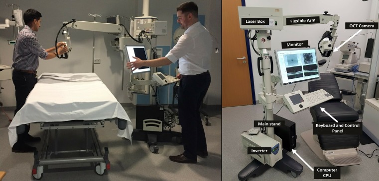
Objective: This study aims to evaluate the feasibility of retinal imaging in critical care using a novel mobile optical coherence tomography (OCT) device. The Heidelberg SPECTRALIS FLEX module (Heidelberg Engineering, Heidelberg, Germany) is an OCT unit with a boom arm, enabling ocular OCT assessment in less mobile patients.
Design: We undertook an evaluation of the feasibility of using the SPECTRALIS FLEX for undertaking ocular OCT images in unconscious and critically ill patients.
Setting: This study was conducted in the critical care unit of a large tertiary referral unit in the United Kingdom.
Participants: 13 systemically unwell patients admitted to the critical care unit were purposively sampled to enable evaluation in patients with a range of clinical states.
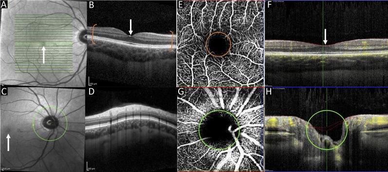
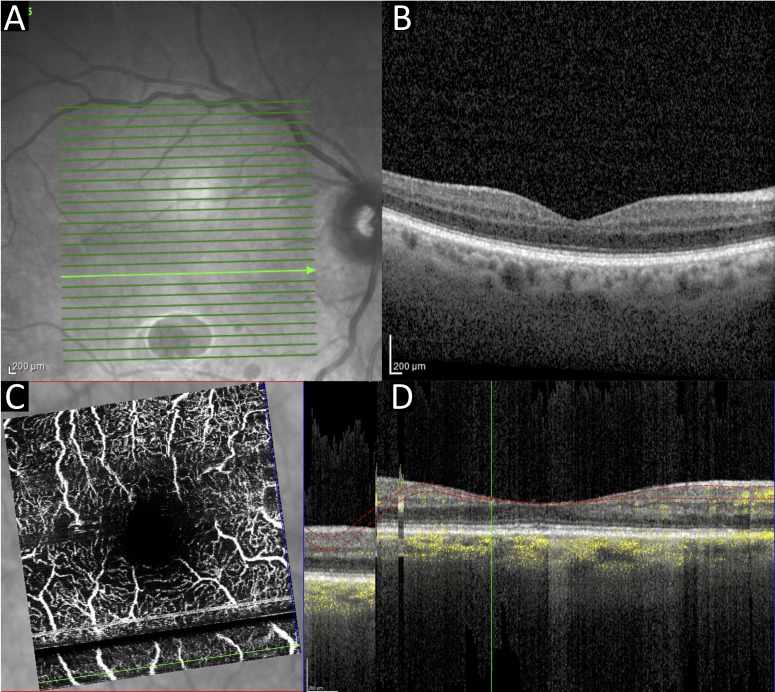
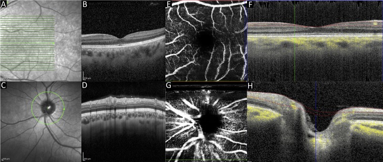
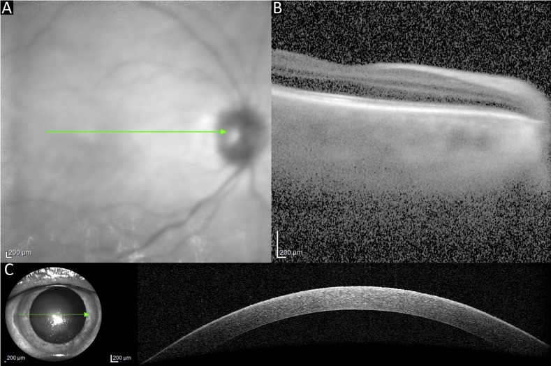
Outcome measures: The primary outcome was the feasibility of acquiring clinically interpretable OCT scans on a consecutive series of patients. The standardised scanning protocol included macula-focused OCT, OCT optic nerve head (ONH), OCT angiography (OCTA) of the macula and ONH OCTA.
Results: OCT images from 13 patients were attempted. The success rates of each scan type are 84% for OCT macula, 76% for OCT ONH, 56% for OCTA macula and 36% for OCTA ONH. The overall mean success rate of scans per patient was 64% (95% CI 46% to 81%). Clinicians reported clinical value in 100% scans which were successfully obtained, including both ruling in and ruling out relevant ocular complications such as corneal thinning, macular oedema and optic disc swelling. The most common causes of failure to achieve clinically interpretable scans were inadequately sustained OCT alignment in delirious patients and a compromised ocular surface due to corneal exposure.
Conclusions: This prospective evaluation indicates the feasibility and potential clinical value of the SPECTRALIS FLEX OCT system on the critical care unit. Portable OCT systems have the potential to bring instrument-based ophthalmic assessment to critically ill patients, enabling detection and micron-level monitoring of ocular complications.
Keywords: adult intensive & critical care; optical coherence tomography; optical coherence tomography angiography.
© Author(s) (or their employer(s)) 2019. Re-use permitted under CC BY-NC. No commercial re-use. See rights and permissions. Published by BMJ.
Conflict of interest statement
Competing interests: None declared.
Figures
References
-
- Kathryn Bigsby BB, et al. Retinal and balance changes based on concussion history: a study of division 1 football players. Int J Phys Med Rehabil 2014;02 10.4172/2329-9096.1000234 - DOI
Publication types
MeSH terms
Grants and funding
LinkOut - more resources
Full Text Sources
Medical