. 2020 Jan 3;295(1):55-68.
doi: 10.1074/jbc.RA119.010902. Epub 2019 Nov 7.

Isolation and structural characterization of a Zn2+-bound single-domain antibody against NorC, a putative multidrug efflux transporter in bacteria

Affiliations

Isolation and structural characterization of a Zn2+-bound single-domain antibody against NorC, a putative multidrug efflux transporter in bacteria

Sushant Kumar et al. J Biol Chem. .

Abstract

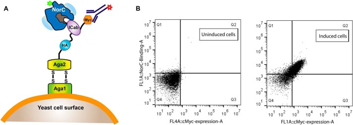
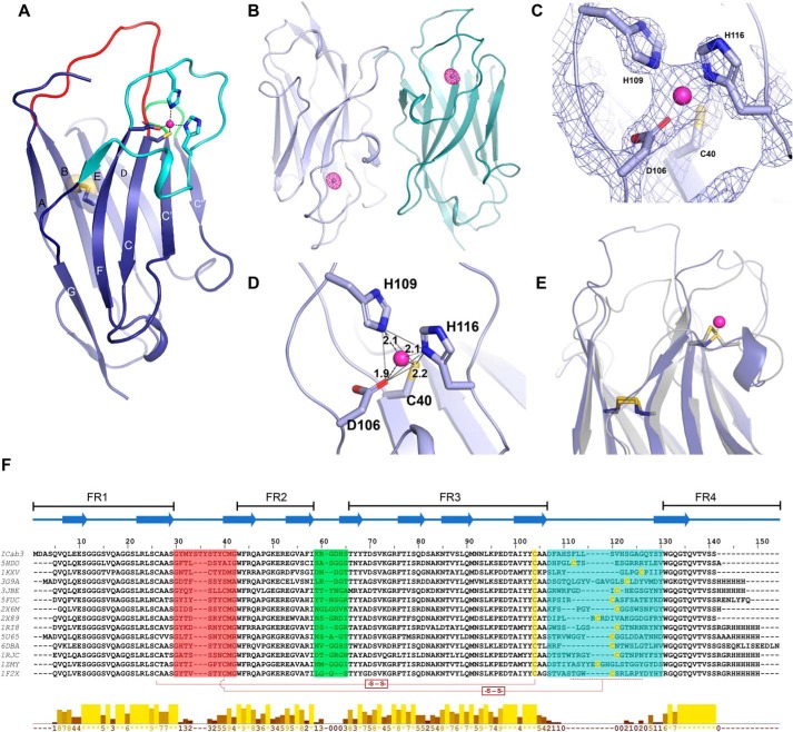
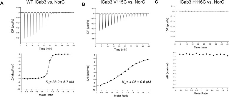
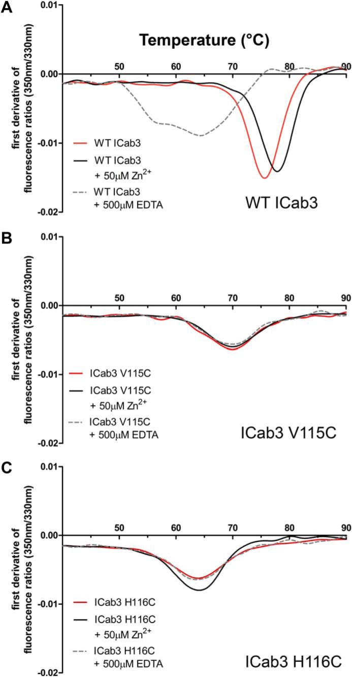
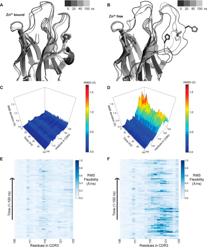
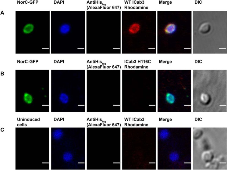
Single-chain antibodies from camelids have served as powerful tools ranging from diagnostics and therapeutics to crystallization chaperones meant to study protein structure and function. In this study, we isolated a single-chain antibody from an Indian dromedary camel (ICab) immunized against a bacterial 14TM helix transporter, NorC, from Staphylococcus aureus We identified this antibody in a yeast display screen built from mononuclear cells isolated from the immunized camel and purified the antibody from Escherichia coli after refolding it from inclusion bodies. The X-ray structure of the antibody at 2.15 Å resolution revealed a unique feature within its CDR3 loop, which harbors a Zn2+-binding site that substitutes for a loop-stabilizing disulfide bond. We performed mutagenesis to compromise the Zn2+-binding site and observed that this change severely hampered antibody stability and its ability to interact with the antigen. The lack of bound Zn2+ also made the CDR3 loop highly flexible, as observed in all-atom simulations. Using confocal imaging of NorC-expressing E. coli spheroplasts, we found that the ICab interacts with the extracellular surface of NorC. This suggests that the ICab could be a valuable tool for detecting methicillin-resistant S. aureus strains that express efflux transporters such as NorC in hospital and community settings.

Keywords: MRSA diagnosis; NorC; X-ray crystallography; antimicrobial efflux; camelid antibody; drug resistance; efflux pump; isothermal titration calorimetry (ITC); major facilitator superfamily (MFS); multidrug transporter; single-domain antibody (sdAb, nanobody); zinc.


Conflict of interest statement

The authors declare that they have no conflicts of interest with the contents of this article

Figures

Figure 1.
Figure 1.
Screening of VHH against NorC using yeast display. A, schematic of the yeast surface display strategy used for screening of VHH (ICab) binders against NorC (T4C) labeled with fluorescein (excitation, 494 nm; emission, 521 nm). A yeast display library built using vhh genes from PBMCs of an immunized camel was screened using anti-c-Myc antibody labeled with Alexa Fluor 647 (excitation, 650 nm; emission, 668 nm). B, FACS screening of uninduced yeast cells displays no shifts when incubated with fluorescein-labeled NorC and displays no nonspecific binding to anti-c-Myc antibody. Induced cells expressing ICab3 display diagonal shifts, signifying binding of NorC to surface-displayed ICab3.
Figure 2.
Figure 2.
Structural features of ICab3. A, overall structure of ICab3. CDR1, CDR2, and CDR3 regions are represented by red, green, and cyan, respectively; transparent spheres depict the disulfide bond between Cys26 and Cys103. B, anomalous map (magenta mesh) of ICab3 contoured at 6.0σ showing a strong density in the CDR3 of each chain. C, 2FoFc map contoured at 3.0σ showing Zn2+-coordinating residues in the Zn2+-binding pocket. D, Zn2+ is bound with a tetrahedral coordination geometry, outlined with gray lines; dashes show distances between Zn2+ and coordinating atoms (Å). E, structural superposition of ICab3 (light blue) with another camelid VHH (gray; PDB code 3JBE) showing overlap of the Zn2+-binding region of ICab3 (light blue) with that of the disulfide bond of 3JBE. F, multiple-sequence alignment of ICab3 with other VHH sequences whose structures are known. β-Strands are represented by overhead arrows, whereas CDR1 to -3 are highlighted in the same color as in A. Cysteines that form disulfides have been shown with connectors (-S-S-), whereas the region in CDR3 that harbors additional cysteine is demarcated with a dashed line. Conservation scores are given in the histogram below. 0, least conservation; *, total conservation. −, unassigned region. FR1–4 represent framework regions 1–4 in the VHH.
Figure 3.
Figure 3.
Binding analysis of ICab3 and its mutants with NorC. A, ITC profile (top, differential power; bottom, binding isotherms with integrated peaks normalized to moles of injectant and offset-corrected) showing nanomolar affinity of WT ICab3 with NorC. B, the binding affinity decreases 100-fold in the case of ICab3 V115C mutant. C, ICab3 H116C has no affinity for NorC.
Figure 4.
Figure 4.
Thermal stability of ICab3 and its mutants. Nano-DSF profiles of WT ICab3 and the mutants showing highest thermal stability for WT ICab3 (A) followed by ICab3 V115C (B) and ICab3 H116C (C). The Tm of WT ICab3 resembles that of ICab3 H116C upon the addition of 0.5 mm EDTA. While, Tm of WT ICab3 increases upon the addition of Zn2+, neither its excess nor the presence of EDTA as its chelator alters the Tm of ICab3 V115C or ICab3 H116C.
Figure 5.
Figure 5.
Molecular dynamics simulations highlighting the role of Zn2+ in providing conformational stability to CDR3. A and B, representation of structure along the trajectory at different time points in the Zn2+-bound (A) and Zn2+-free (B) state. In the presence of Zn2+, CDR3 loop conformation is restrained, whereas in the absence of Zn2+, it becomes more mobile, which allows the His residue (involved in coordination to Zn2+) to flip outside. C and D, RMSD of residues in the CDR3 region over the course of trajectory in the presence of Zn2+ (C) and in the absence of Zn2+ (D). E and F, RMS flexibility of residues in the CDR3 region in Å/ns in the presence of Zn2+ (E) and in the absence of Zn2+ (F); only side-chain atoms were taken for calculation. Conformational flexibility of residues from 115 to 121 significantly increases in the absence of Zn2+ coordination.
Figure 6.
Figure 6.
Confocal imaging showing WT ICab3 binding to the extracellular face of NorC in E. coli spheroplasts. Images were acquired after incubating E. coli spheroplasts expressing NorC-GFP with rhodamine-labeled WT ICab3 (A) and rhodamine-labeled ICab3 H116C (B). C, images acquired with uninduced E. coli spheroplasts incubated with rhodamine-labeled WT ICab3. Total number of cells counted for A = 40, and that for B and C was 15 each in four trials. Scale bar, 1 μm. DIC, differential interference contrast.

References

    1. Walsh C. (2000) Molecular mechanisms that confer antibacterial drug resistance. Nature 406, 775–781 10.1038/35021219 - DOI - PubMed
    1. Webber M. A., and Piddock L. J. (2003) The importance of efflux pumps in bacterial antibiotic resistance. J. Antimicrob. Chemother. 51, 9–11 10.1093/jac/dkg050 - DOI - PubMed
    1. Reddy V. S., Shlykov M. A., Castillo R., Sun E. I., and Saier M. H. Jr. (2012) The major facilitator superfamily (MFS) revisited. FEBS J. 279, 2022–2035 10.1111/j.1742-4658.2012.08588.x - DOI - PMC - PubMed
    1. Saier M. H. Jr., Reddy V. S., Tamang D. G., and Västermark A. (2014) The transporter classification database. Nucleic Acids Res. 42, D251–D258 10.1093/nar/gkt1097 - DOI - PMC - PubMed
    1. Paulsen I. T., Brown M. H., and Skurray R. A. (1996) Proton-dependent multidrug efflux systems. Microbiol. Rev. 60, 575–608 - PMC - PubMed

Publication types

MeSH terms

LinkOut - more resources