An Unusual Case of Left Spigelian Hernia Containing Part of a Fibroid Uterus and the Left Adnexa in a 66-Year-Old Woman
- PMID: 31699961
- PMCID: PMC12560673
- DOI: 10.12659/AJCR.917104
An Unusual Case of Left Spigelian Hernia Containing Part of a Fibroid Uterus and the Left Adnexa in a 66-Year-Old Woman
Abstract
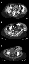
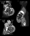
BACKGROUND Spigelian hernia, or lateral ventral hernia, is rare and represents between 0.1-2% of all hernias of the abdominal wall. The presentation is variable, and the diagnosis may be challenging. This report is of an unusual case of Spigelian hernia that contained part of a fibroid uterus and the left adnexa. CASE REPORT A 66-year-old woman presented with an abdominal wall mass in the left lower quadrant. On physical examination, a provisional diagnosis of ventral hernia was made. Abdominal computed tomography (CT) imaging showed an unusual Spigelian hernia that contained part of a fibroid uterus and the left adnexa. Treatment using laparoscopic hysterectomy, left salpingo-oophorectomy, and hernia repair was successfully performed jointly by a general surgeon and a gynecologist. CONCLUSIONS To the best of our knowledge, the is the first reported case of Spigelian hernia that contained part of the uterus and the left adnexa.
Conflict of interest statement
Figures


References
-
- Park AE, Roth JS, Kavic SM. Abdominal wall hernia. Curr Probl Surg. 2006;43(5):326–75. - PubMed
-
- Houlihan TJ. A review of Spigelian hernias. Am J Surg. 1976;131(6):734–35. - PubMed
-
- Moles Morenilla L, Docobo Durántez F, Mena Robles J, de Quinta Frutos R. Spigelian hernia in Spain: An analysis of 162 cases. Rev Esp Enferm Dig. 2005;97(5):338–47. - PubMed
-
- Bhatia TP, Ghimire P, Panhani ML. Spigelian hernia. Kathmandu Univ Med J. 2010;8(30):241–43. - PubMed
Publication types
MeSH terms
LinkOut - more resources
Full Text Sources

