. 2019 Nov 7;9(1):16241.
doi: 10.1038/s41598-019-52335-x.

Cellular elements in the developing caecum of Japanese quail (Coturnix coturnix japonica): morphological, morphometrical, immunohistochemical and electron-microscopic studies

Affiliations

Cellular elements in the developing caecum of Japanese quail (Coturnix coturnix japonica): morphological, morphometrical, immunohistochemical and electron-microscopic studies

Aalaa M AbuAli et al. Sci Rep. .

Abstract

The present study aims to investigate the histological, histochemical and electron microscopic changes of the caecal proximal part of Japanese quail during both pre- and post-hatching periods starting from the 2nd embryonic day (ED) until four weeks post-hatching. On the 2nd and 3rd ED, the primordia of caeca appeared as bilateral swelling on the wall of the hindgut. On the 7th ED, the lamina propria/submucosa contained the primordia of glands. On the 8th ED, rodlet cells could be observed amongst the epithelial cells. On the 9th ED, the caeca began to divide into three parts with more developed layers. With age, the height and number of villi increased. On the 13th ED, immature microfold cells (M-cells) could be identified between the surface epithelium of the villi. The caecal tonsils (CTs) appeared in the form of aggregations of lymphocytes, macrophages, dendritic cells and different types of leukocytes. Telocytes and crypts of Lieberkuhn were observed at this age. On hatching day, the crypts of Lieberkuhn were well-defined and formed of low columnar epithelium, goblet cells, and enteroendocrine cells. Post-hatching, the lumen was filled with villi that exhibited two forms: (1) tongue-shaped villi with tonsils and (2) finger-shaped ones without tonsils. The villi lining epithelium contained simple columnar cells with microvilli that were dispersed with many goblet cells, in addition to the presence of a high number of intra-epithelial lymphocytes and basophils. Moreover, the submucosa was infiltrated by numerous immune cells. CD3 immunomarker was expressed in intraepithelial lymphocytes, while CD20 immunomarker showed focal positivity in CTs. In conclusion, the caecal immune structures of quails at post-hatching were more developed than those in pre-hatching life. The high frequency of immune cells suggests that this proximal part may be a site for immunological surveillance in the quail caecum. The cellular organisation of the caecum and its relation to the immunity was discussed.


Conflict of interest statement

The authors declare no competing interests.

Figures
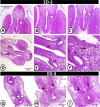
Figure 1
Figure 1
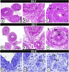
Transverse section (T.S) in the primitive hindgut on the 2nd and 3rd embryonic day (ED) stained by Haematoxylin and Eosin (H&E). (A,B) The beginning of invagination in the hindgut mucosa (arrowhead). (C) The complete invagination of caeca (arrowhead). (D) Primordia of caeca between mesonephros (Ms) and spinal cord (Sc). (E) Primordia of caeca (pC) between the hindgut wall (HG). (F) Hindgut mucosa (HG), undifferentiated mesenchymal tissue (Mes) and mucosa of caeca (M). (G) The hindgut tube showed invagination of caeca (arrowhead). (H) Partial separation of hindgut parts (asterisk). (I) The beginning of caecal tube formation (asterisk).
Figure 2
Figure 2
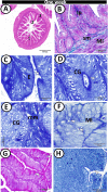
T.S in the caecum on the 5th, 7th, 8th ED, (A-F) stained by H&E, (G-I) stained by toluidine blue. (A,B) Partial separation of the serosal layer (arrowheads) of caeca (C) and ileum (I). (C) The epithelium of caecal mucosa (M) showing mitotic divisions (asterisk), followed by lamina propria/submucosa (arrowhead), muscle layer (Ml) and serosa (S). (D) The ileum (I) between the two caeca (C). (E) Higher magnification showed the incomplete separation of serosa (arrowhead). (F) The caecal wall consisted of mucosa (M), submucosa (sm) with the primordia of the caecal glands (*), the muscle layer (Ml) and serosa (S). (G) Semithin section showed mucosal epithelium (E) with rodlet cells (arrowheads). (H,I) Submucosa contained macrophage (arrowhead) and primordia of caecal glands (CG) with mitotic divisions (asterisk).
Figure 3
Figure 3
T.S in the proximal part of caeca on the 9th and 11th ED. (A,B) The beginning of the proximal part of caeca (C), the ileum (I) and the caecal layers: mucosa (M), submucosa (sm), and muscle layer (Ml) stained by HE. (C) PAS-positive epithelial microvilli (arrowhead). (D) The proximal part of the caecum (C) and the ileum (I). (E) The proximal part layers: mucosa (M), lamina propria (Lp) and muscle layer (Ml). (F) The proximal part showed PAS-positive brush borders (arrowhead).
Figure 4
Figure 4
T.S in the proximal part of caeca on the 13th and 15th ED. (A) Two shapes of mucosal villi stained by HE; branched finger-shaped villi (1), finger-shaped villi (2). (B) Semithin section stained by toluidine blue showed a part of villi occupied with lamina propria (Lp) and covered by simple epithelium (E) and rodlet cells (arrowheads). (C,D) Submucosa with crypts of Lieberkuhn (asterisks). (E) Bromophenol blue-positive rodlet cell (RC) and brush borders (arrowhead). (F) PAS-positive goblet cell (gc) and brush borders (arrowhead). (G) General view of the proximal part of caeca (C) and ileum (I) stained by HE. (H) The wall of the proximal part showed mucosal folds (Mf), mucosa (M) and muscle layer (Ml). (I) The mucosal folds showed PAS-positive goblet cells (arrowheads) and microvilli (asterisk).
Figure 5
Figure 5
T.S in the proximal part of caeca at hatching day. (A) Two shapes of villi (1&2). (B) The finger-shaped villi filled with lamina propria (Lp) and covered by epithelium (E) with goblet cell (arrowhead). (C) PAS-positive goblet cells (arrowheads) and brush borders (asterisk). (D) Semithin section stained by toluidine blue (TB) showed the surface epithelium (E) dispersed by goblet cells (asterisk). Note, telocytes (arrowhead) in association with blood capillaries (BC). (E,F) Lamina propria showed crypts of Lieberkuhn (CL), and caecal tonsils (CT). (G) Telocytes (arrowheads) surrounded crypts of Lieberkuhn (CL) with endocrine cells (asterisks). (H,I) Muscle layer (Ml) infiltrated by nerve bundles (asterisks) and telocytes (arrowheads).
Figure 6
Figure 6
T.S in the proximal part of caeca at one-week post-hatching. (A) Shapes of mucosal folds; finger 1, leaf-like 2 & tongue shape 3. (B) The wall of the caecum stained by Crossmon’s Trichrome showing muscle layer (Ml), submucosa (sm) and lamina propria (Lp). (C-E) Semithin section showed the epithelium (E) of the mucosal fold and caecal glands (CG) with prominent microvilli (arrowhead), surrounded by muscularis mucosa (mm). (F) Outer and inner muscle layers (Ml) separated by fat cells (arrowheads). (G) PAS-positive goblet cells (arrowheads). (H) Bromophenol blue-positive apical epithelial surface (arrowhead).
Figure 7
Figure 7
T.S in the proximal part of caeca at two weeks post-hatching. (A,B) Many finger-shaped villi (MF) covered by simple epithelium (E). (C,D) The lamina propria contained crypts of Lieberkuhn (CL) and caecal tonsils (CT) (arrowhead, boxed areas). (E) The villi filled with lamina propria (Lp) and covered by epithelium (E) infiltrated by goblet cells (arrowheads) and intraepithelial lymphocytes (IELs) (asterisk). (F) PAS-positive goblet cells (arrowheads). (G) Bromophenol blue-positive brush borders and apical portion of the crypts of Lieberkuhn (arrowheads).
Figure 8
Figure 8
Immunohistochemical expression pattern of CD3 and CD20 in the proximal part of caeca at hatching day. (A) Intraepithelial lymphocytes (arrowheads) expressed immunopostivity to CD3. (B) Lamina propria showed CD3-immunopositive T-lymphocytes (boxed areas) around the crypts of Lieberkuhn (CL). (C,D) CD20-positive B-lymphocytes were immunolocalised in the caecal tonsils (boxed area). Note, the negative control (boxed area) in A,C.
Figure 9
Figure 9
Immunohistochemical expression pattern of Secretory immunoglobulin A (SIgA) and s100 protein in the proximal part of caeca at hatching day. (A,B) SIgA-positive cells (arrowheads) in the lamina propria (LP) under the epithelium (EP) and the crypts of Lieberkuhn (CL). (C) Positive-SIgA in the epithelial cells (arrowheads). Note, an intense staining reaction in the outer epithelial membrane (arrow). (D) S-100 protein-immunopositive dendritic cells (boxed area) in the lamina propria (LP). (E,F) S-100 protein-positive telocytes (arrowheads, boxed areas) between the muscle layers. Note, the negative control (boxed area) in A,D.
Figure 10
Figure 10
Scanning electron microscopy (SEM) of the caeca on the 8th ED. (A) The surface epithelium covered by individual cilium (arrowheads). (B) Surface view showing flattened polyhedral epithelium (E) with serrated edges (arrowhead). (C,D) Lateral view showing the epithelium (E) and honeycomb-shaped villi.
Figure 11
Figure 11
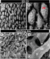
SEM of the proximal part of caeca on the 13th ED and day of hatching. (A,B) Mucosal folds (Mf) with honeycomb villus surface. Note deep cavities of crypts of Lieberkuhn (arrowhead). (C,D) Many mucosal folds (MF) with pores for crypts of Lieberkuhn (arrowheads).
Figure 12
Figure 12
SEM of the proximal part of caeca at one and two weeks post-hatching. (AC) The caecal villi (V) covered by cilia (asterisk). Note, pores of goblet cells (arrowhead). (D,E) A high number of the villi (V) in two-weeks quail showed cilia (asterisk) and numerous pores of goblet cells (arrowhead). (F) The surface epithelium (E) covered by individual cilium (arrowheads).
Figure 13
Figure 13
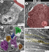
Digital coloured transmission electron microscopic (TEM) images of the proximal part of caeca on the 13th ED. (A,B) The epithelium consisted of simple columnar cells (yellow colour) with brush borders (red colour), and connected to the neighbouring cells by desmosomes (arrowheads). Note M-cell (M, pink colour), and the infiltration of the epithelium by dendritic cell (green) and lymphocytes (L, violet). (C,D) Goblet cells (blue colour), and rodlet cell (green colour) are distributed in the epithelium.
Figure 14
Figure 14
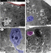
Digital coloured TEM images of caecum on the 13th ED. (A) Surface epithelium with euchromatic nucleus (N), contained rough endoplasmic reticulum (R) and mitochondria (M) and connected by desmosomes (arrowheads). (B) The crypts of Lieberkuhn (red colour) are surrounded by smooth muscle cells (arrowheads). (C) Submucosa contained macrophages (pink, Mc), lymphocytes (orange, L), dendritic cell (green, D), and fibroblast (blue, F). (D) Muscle layer infiltrated by telocytes (TC) and their telopodes (Tp) (yellow colour).
Figure 15
Figure 15
Digital coloured TEM images of caecum at hatching day. (A) The epithelium connected by desmosomes (arrowheads), covered by microvilli (red), and the cytoplasm contained rough endoplasmic reticulum (R) and mitochondria (M). (B,C) Surface epithelium with neuroendocrine cell (blue colour). (D) Intraepithelial lymphocyte (pink) and basophile leukocyte (dark colour) are migrated to the epithelium.
Figure 16
Figure 16
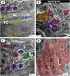
Digital coloured TEM images of caecum at hatching day. (AC) Submucosa contained clusters of lymphocytes (violet, L), dendritic cell (pink, DC), Macrophages (orange, Mc), developing leukocytes (green, DL) and blood capillaries (BC) surrounded by telocytes (yellow, TC) and fibroblast (blue, F). (D) Crypts of Lieberkuhn (red) with enteroendocrine cells contained electron-dense granules (arrowhead).
Figure 17
Figure 17
Digital coloured TEM images of caecum at one-week post-hatching. (A-C) Simple epithelium contained rough endoplasmic reticulum (R), mitochondria (M), dispersed by goblet cells (blue colour, asterisks), connected with desmosome (arrowheads) and covered by microvilli (red colour). (D) Bundles of nerve fibres (N) between the muscles.
Figure 18
Figure 18
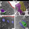
Digital coloured TEM images of caecum at one-week post-hatching. (A,B) Enteroendocrine cells with dense granules (purple colour), goblet cells (blue colour) and telocytes (yellow colour) under the basement membrane. (C,D) telocytes (yellow) and macrophage (green) in the submucosa.

Similar articles

Cited by

References

    1. Padgett CS, Ivey WD. The normal embryology of the Coturnix quail. Anat. Rec. 1960;137:1–11. doi: 10.1002/ar.1091370102. - DOI - PubMed
    1. Zacchei AM. The embryonal development of the Japanese quail (Coturnix coturnix japonica) Arch. Ital. Anat. Embryol. 1961;66:36–62. - PubMed
    1. Hamburger V, Hamilton HL. A series of normal stages in the development of the chick embryo. Devel. Dyn. 1992;195:229–30. doi: 10.1002/aja.1001950403. - DOI - PubMed
    1. Ainsworth SJ, Stanley RL, Evans DJR. Developmental stages of the Japanese quail. J. Anat. 2010;216:3–15. doi: 10.1111/j.1469-7580.2009.01173.x. - DOI - PMC - PubMed
    1. Huss D, Poynter G, Lansford R. Japanese quail (Coturnix japonica) as a laboratory animal model. Lab. Anim. 2008;37:513–519. doi: 10.1038/laban1108-513. - DOI - PubMed

Publication types