. 2019 Nov 7;9(1):16178.
doi: 10.1038/s41598-019-52824-z.

Direct interaction between the hepatitis B virus core and envelope proteins analyzed in a cellular context

Affiliations

Direct interaction between the hepatitis B virus core and envelope proteins analyzed in a cellular context

Florentin Pastor et al. Sci Rep. .

Abstract

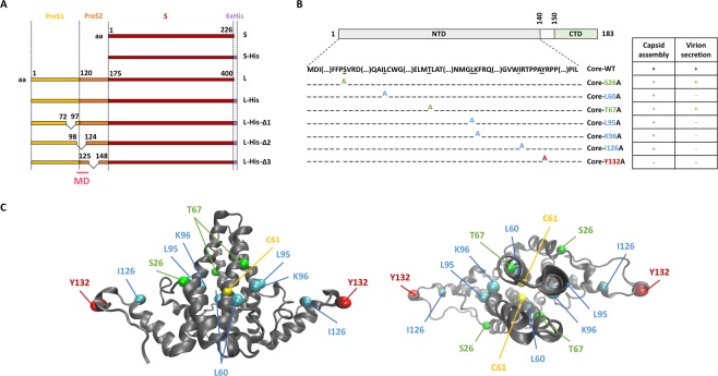
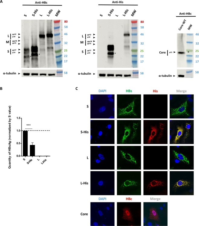
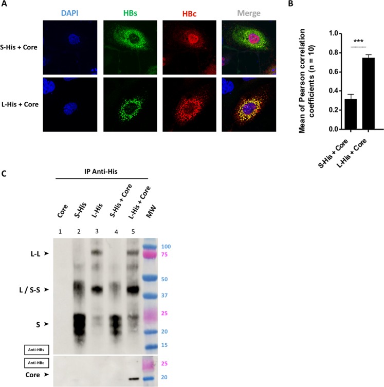
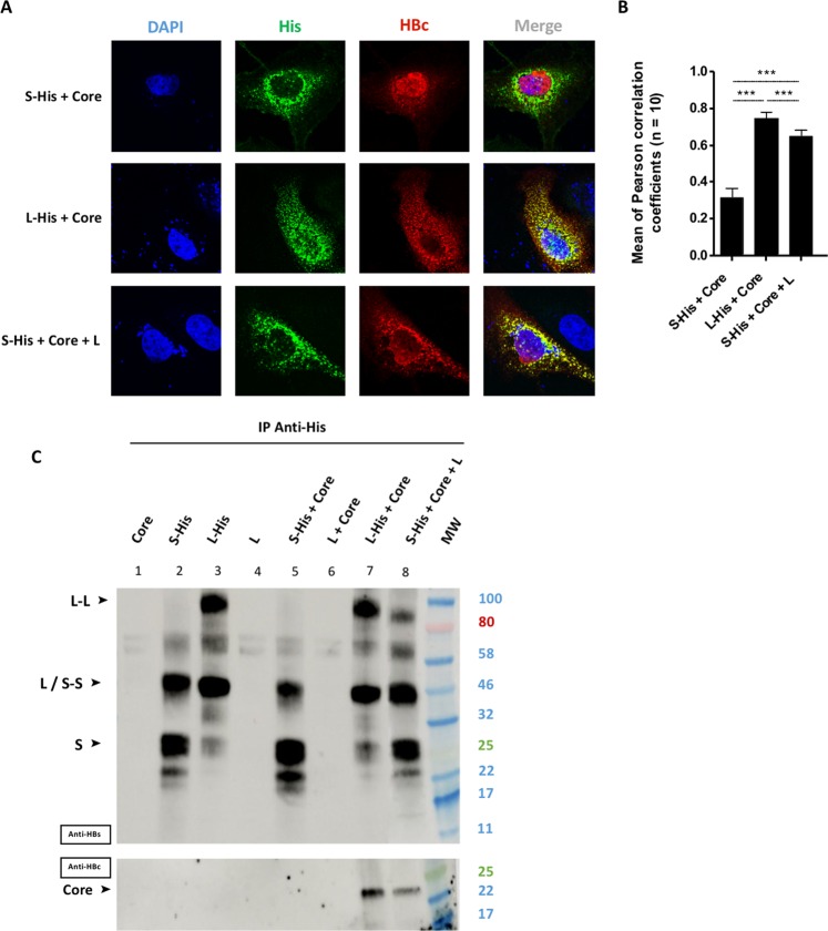
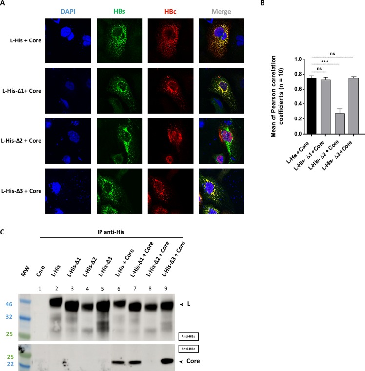
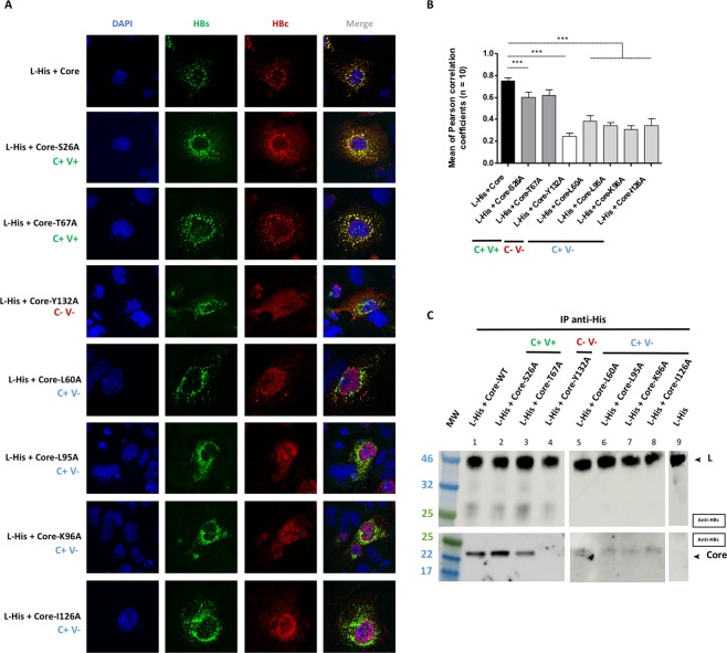
Hepatitis B virus (HBV) production requires intricate interactions between the envelope and core proteins. Analyses of mutants of these proteins have made it possible to map regions involved in the formation and secretion of virions. Tests of binding between core and envelope peptides have also been performed in cell-free conditions, to study the interactions potentially underlying these mechanisms. We investigated the residues essential for core-envelope interaction in a cellular context in more detail, by transiently producing mutant or wild-type L, S, or core proteins separately or in combination, in Huh7 cells. The colocalization and interaction of these proteins were studied by confocal microscopy and co-immunoprecipitation, respectively. The L protein was shown to constitute a molecular platform for the recruitment of S and core proteins in a perinuclear environment. Several core amino acids were found to be essential for direct interaction with L, including residue Y132, known to be crucial for capsid formation, and residues L60, L95, K96 and I126. Our results confirm the key role of L in the tripartite core-S-L interaction and identify the residues involved in direct core-L interaction. This model may be valuable for studies of the potential of drugs to inhibit HBV core-envelope interaction.


Conflict of interest statement

The authors declare no competing interests.

Figures

Figure 1
Figure 1
Diagram of the S, L and core proteins and their mutated derivatives. (A) The WT S envelope protein consists of the S domain (red) and the WT L envelope protein consists of this domain together with two other domains at its N-terminus: the preS1 (yellow) and the preS2 (orange) domains. The matrix domain (MD) is indicated (pink). Deletions are indicated by a bridge linking the flanking residues. Some constructs contain a His-tag at their C-terminus (purple box). (B) The WT core protein consists of the NTD domain (light gray), a linker (white) and the CTD domain (green). Seven mutant core proteins were generated by alanine substitution. The ability of mutated core proteins to assemble and to produce virions, according to the findings of Ponsel et al., is summarized in the table on the right. The names of the constructs are shown in color, according to their capsid assembly and virion secretion properties: C+V+ residues in green, C+V− residues in blue, C-V- residues in red. (C) Orthogonal views of the 3D structure of the core NTD dimer linked by C61 residues (in yellow). This structure was obtained by crystallography (#1QGT). The backbone is represented as a gray ribbon. Mutated residues are shown in the CPK model, with colors as in the table in (B).
Figure 2
Figure 2
Envelope and core protein levels. Huh7 cells were transfected with a plasmid encoding the untagged WT HBV S protein or S-His, or WT untagged L, L-His or core proteins. Three days after transfection, the cells were analyzed by western blotting for HBsAg secretion, and by confocal microscopy. (A) Cell lysates were separated by SDS-PAGE, the bands were transferred onto membranes and the membranes were probed with anti-HBs (left panel), anti-His (central panel) or anti-HBc (right panel) antibodies. The asterisk (*) notes the presence of a truncated version of the S and S-His proteins. (B) A commercial ELISA was used to quantify HBsAg in cell supernatants. The amount of S-His secreted was about half the amount of WT S secreted. The bars indicate the mean ± standard deviation (SD) values from four independent experiments. p values (paired t-tests) were determined: ***p value < 0.001. (C) Cells were fixed on coverslips and proteins were visualized by confocal microscopy after indirect immunofluorescence with an anti-HBs antibody (in green) together with an anti-His antibody (in red) for the L or S proteins, or with an anti-HBc antibody (in red) for the core protein. Nuclei were labeled with DAPI (in blue).
Figure 3
Figure 3
Only the L protein interacts with the core protein. Huh7 cells were cotransfected by incubation for three days with a plasmid encoding the S-His or L-His protein together with a plasmid encoding the core protein. (A) Huh7 cells were fixed and double-stained with anti-HBs (in green) and anti-HBc (in red) antibodies, and the nuclei were counterstained with DAPI (in blue). Each image corresponds to the major phenotype observed. Note that the colocalization of envelope and core proteins was observed only for the L-His and core proteins. (B) Histogram of r values. Differences between L-His/Core and S-His/Core colocalizations were analyzed statistically by Mann-Whitney tests on 30 images from 3 different experiments (***p < 0.001). (C) Huh7 cell lysates were collected and 400 µg of total protein was subjected to IP with an anti-His antibody. Membranes were probed with an anti-HBs (IP) or an anti-HBc (Co-IP) antibody. Note that the co-IP of core protein was observed only in the presence of L-His protein.
Figure 4
Figure 4
Tripartite interaction between the L, S and core proteins. Huh7 cells were cotransfected with a plasmid encoding the core protein, and a plasmid encoding S-His, or L-His, or S-His plus untagged L. Three days post-transfection, cells were analyzed by confocal microscopy and co-IP. (A) Cells were fixed and double-stained with anti-His (in green) and anti-HBc (in red) antibodies, and nuclei were labeled with DAPI (in blue). S-His was colocalized with WT core protein in the presence of untagged L proteins (bottom row). (B) Histogram of r values (***p value < 0.001). (C) Huh7 cell lysates were collected and 400 µg of total protein was subjected to IP with an anti-His antibody. The immunoprecipitated samples were blotted and the membranes were probed with anti-HBs (IP) or anti-HBc (Co-IP) antibodies. Core protein co-IP was observed with S-His if untagged L was also produced.
Figure 5
Figure 5
Interaction between the mutant L proteins and the core protein. Huh7 cells were cotransfected with plasmids encoding the L-His, L-His-Δ1, L-His-Δ2 or L-His-Δ3 protein and a plasmid encoding the core protein. Three days post-transfection, cells were analyzed by confocal microscopy and co-IP. (A) Cells were fixed and double-stained with anti-HBs (in green) and anti-HBc (in red) antibodies, and nuclei were labeled with DAPI (in blue). Perfect colocalization with the core protein was observed for the L-His protein, and the L-His-Δ1 and L-His-Δ3 mutant proteins, but not for the L-His-Δ2 mutant. (B) Histogram of r values as previously described (***p value < 0.001). (C) Huh7 cell lysates were collected and 400 µg of total protein was subjected to IP with an anti-His antibody. Membranes were probed with anti-HBs (IP) or anti-HBc (Co-IP) antibodies. Well 1 is a control, demonstrating a lack of core protein immunoprecipitation with the anti-His antibody. Wells 2–5 are controls showing that L-His and L-His derivatives are immunoprecipitated with the anti-His antibody. Core protein co-IP was observed with L-His and all derivatives except for the L-His-Δ2 mutant.
Figure 6
Figure 6
Interaction between the L protein and the mutant core proteins. Huh7 cells were cotransfected with plasmids encoding the L-His protein and the WT core protein or one of the seven mutant core proteins. Three days post-transfection, cells were analyzed by immunolabeling and confocal microscopy, and co-IP was investigated with L-His as the bait. (A) Cells were double-stained with anti-HBs (in green) and anti-HBc (in red) antibodies, and nuclei were labeled with DAPI (in blue). As for the WT controls, a strong colocalization of the L-His and core proteins was observed with C+V+ mutants (core-S26A and coreT67A). Partial colocalization between the L-His and core proteins was observed with C+V− mutants (core-L60A, core-L95A, core-K96A, core-I126A) and no colocalization of L-His and core proteins was observed for the C-V- mutant (core-Y132A). (B) Histogram of r values, as previously described (***p value < 0.001). The capsid formation and secretion properties of each mutant are shown at the bottom of the figure. (C) Huh7 cell lysates were collected and 400 µg of total protein was subjected to IP with an anti-His antibody. The immunoprecipitated samples were subjected to immunoblotting and the membranes were probed with anti-HBs (IP) or anti-HBc (Co-IP) antibodies. The capsid formation and virion secretion properties of each of the mutant proteins are reported at the top of the figure. The C+V+ mutants were strongly co-immunoprecipitated with the WT L-His. Weak but detectable co-IP was observed with the C+V− mutants, whereas no signal was obtained with the C−V− mutant, as for the controls (simple transfection of each partner) (data not shown). All the blot acquisitions were processed in parallel and correspond to the same experiment.

References

    1. Trepo C. A brief history of hepatitis milestones. Liver Int. 2014;34(Suppl 1):29–37. doi: 10.1111/liv.12409. - DOI - PubMed
    1. Revill PA, et al. A global scientific strategy to cure hepatitis B. Lancet. Gastroenterol Hepatol. 2019;4:545–558. - PMC - PubMed
    1. Seeger C, Mason WS. Molecular biology of hepatitis B virus infection. Virology. 2015;479–480:672–686. doi: 10.1016/j.virol.2015.02.031. - DOI - PMC - PubMed
    1. Summers J, Mason WS. Replication of the genome of a hepatitis B–like virus by reverse transcription of an RNA intermediate. Cell. 1982;29:403–415. doi: 10.1016/0092-8674(82)90157-X. - DOI - PubMed
    1. Hu, J. & Liu, K. Complete and Incomplete Hepatitis B Virus Particles: Formation, Function, and Application. Viruses9 (2017). - PMC - PubMed

Publication types

MeSH terms

Substances