. 2019 Nov 7;9(1):16278.
doi: 10.1038/s41598-019-52637-0.

Computational and NMR studies of RNA duplexes with an internal pseudouridine-adenosine base pair

Affiliations

Computational and NMR studies of RNA duplexes with an internal pseudouridine-adenosine base pair

Indrajit Deb et al. Sci Rep. .

Abstract

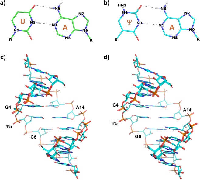
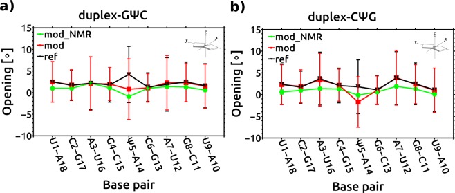
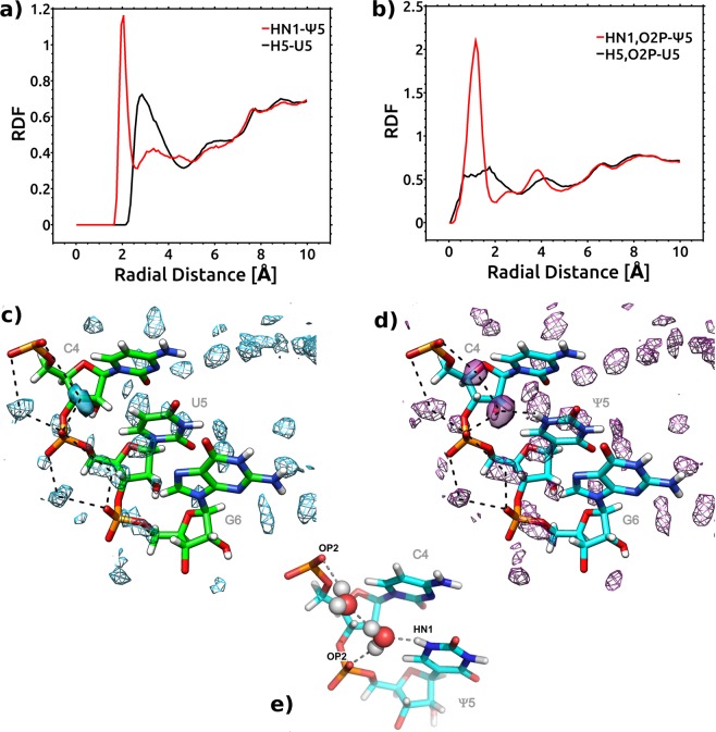
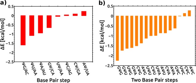
Pseudouridine (Ψ) is the most common chemical modification present in RNA. In general, Ψ increases the thermodynamic stability of RNA. However, the degree of stabilization depends on the sequence and structural context. To explain experimentally observed sequence dependence of the effect of Ψ on the thermodynamic stability of RNA duplexes, we investigated the structure, dynamics and hydration of RNA duplexes with an internal Ψ-A base pair in different nearest-neighbor sequence contexts. The structures of two RNA duplexes containing 5'-GΨC/3'-CAG and 5'-CΨG/3'-GAC motifs were determined using NMR spectroscopy. To gain insight into the effect of Ψ on duplex dynamics and hydration, we performed molecular dynamics (MD) simulations of RNA duplexes with 5'-GΨC/3'-CAG, 5'-CΨG/3'-GAC, 5'-AΨU/3'-UAA and 5'-UΨA/3'-AAU motifs and their unmodified counterparts. Our results showed a subtle impact from Ψ modification on the structure and dynamics of the RNA duplexes studied. The MD simulations confirmed the change in hydration pattern when U is replaced with Ψ. Quantum chemical calculations showed that the replacement of U with Ψ affected the intrinsic stacking energies at the base pair steps depending on the sequence context. The calculated intrinsic stacking energies help to explain the experimentally observed sequence dependent changes in the duplex stability from Ψ modification.


Conflict of interest statement

The authors declare no competing interests.

Figures

Figure 1
Figure 1
Chemical structure of (a) uridine and (b) pseudouridine.
Figure 2
Figure 2
Fingerprint (H6/H8-H1′) regions of the 400 ms NOESY spectra recorded in D2O at 25 °C. The sequential connectivities are traced with either a blue or a green line for both strands of the (a) duplex-GΨC and (b) duplex-CΨG.
Figure 3
Figure 3
Base pairing of (a) uridine and (b) pseudouridine with adenosine. Superposition of the ten lowest-energy solution NMR structures: (c) duplex-GΨC, (d) duplex-CΨG.
Figure 4
Figure 4
Average opening values (deg) for the base pairs in (a) duplex-GΨC and (b) duplex CΨG. Green – Ψ-modified NMR models; Red – Ψ-modified duplexes from MD simulation; Black – reference, unmodified from MD simulation. Vertical lines represent standard deviations.
Figure 5
Figure 5
Change in the hydration pattern upon U to Ψ modification for duplex-CΨG and duplex-CUG. RDFs of water oxygen atoms (a) around the HN1-Ψ5 and H5-U5 atoms, respectively; (b) around the geometric center of the OP2-Ψ5 and HN1-Ψ5 atoms or OP2-U5 and H5-U5 atoms. Red – Ψ-modified duplexes; Black – unmodified duplexes. Water occupancy contoured at equivalent levels and hydrogen bonding patterns (in dotted lines) viewed from the major groove. (c) Reference duplex-CUG; (d) Ψ-modified duplex-CΨG; differences in hydration patterns are shown in solid. (e) Snapshot of two water molecules making contact between HN1, OP2 atoms of Ψ and OP2 atom of the preceding residue.
Figure 6
Figure 6
Impact of Ψ on the intrinsic stacking energies. (a) Change in the QM stacking energy between base pairs at a given base pair step upon Ψ modification (ΔE = Emodif − Eunmodif). ΨG/AC denotes the 5′ΨG/3′AC base pair step and so on. (b) Prediction of the impact of Ψ on the stacking energies at trinucleotide steps based on QM calculations. GΨG denotes the 5′GΨG/3′CAC motif and so on. The base pair stacking energies at trinucleotide steps were calculated by the sum of the base pair stacking energies for two consecutive dinucleotide steps. The data are ordered in decreasing stability of eight unique dinucleotide steps/ sixteen unique trinucleotide steps in RNA duplexes.

References

    1. Davis FF, Allen FW. Ribonucleic acids from yeast which contain a fifth nucleotide. J. Biol. Chem. 1957;227:907–915. - PubMed
    1. Gray MCMW. Pseudouridine in RNA: What, Where, How, and Why. I. UBMB Life (International Union Biochem. Mol. Biol. Life) 2000;49:341–351. doi: 10.1080/152165400410182. - DOI - PubMed
    1. Carlile TM, et al. Pseudouridine profiling reveals regulated mRNA pseudouridylation in yeast and human cells. Nature. 2014;515:143–146. doi: 10.1038/nature13802. - DOI - PMC - PubMed
    1. Schwartz S, et al. Transcriptome-wide mapping reveals widespread dynamic-regulated pseudouridylation of ncRNA and mRNA. Cell. 2014;159:148–162. doi: 10.1016/j.cell.2014.08.028. - DOI - PMC - PubMed
    1. Li X, et al. Chemical pulldown reveals dynamic pseudouridylation of the mammalian transcriptome. Nat. Chem. Biol. 2015;11:592–597. doi: 10.1038/nchembio.1836. - DOI - PubMed

Publication types