. 2019 Nov;25(11):1728-1732.
doi: 10.1038/s41591-019-0626-9. Epub 2019 Nov 7.

Genomic and epidemiological evidence of bacterial transmission from probiotic capsule to blood in ICU patients

Affiliations

Genomic and epidemiological evidence of bacterial transmission from probiotic capsule to blood in ICU patients

Idan Yelin et al. Nat Med. 2019 Nov.

Abstract

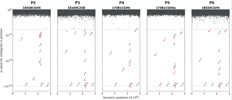
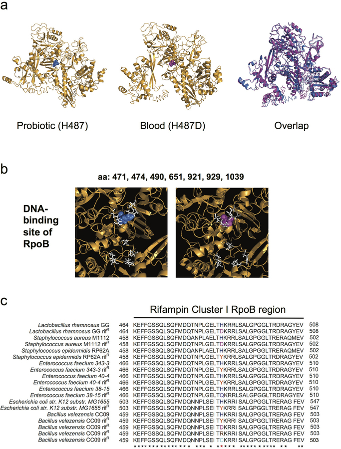
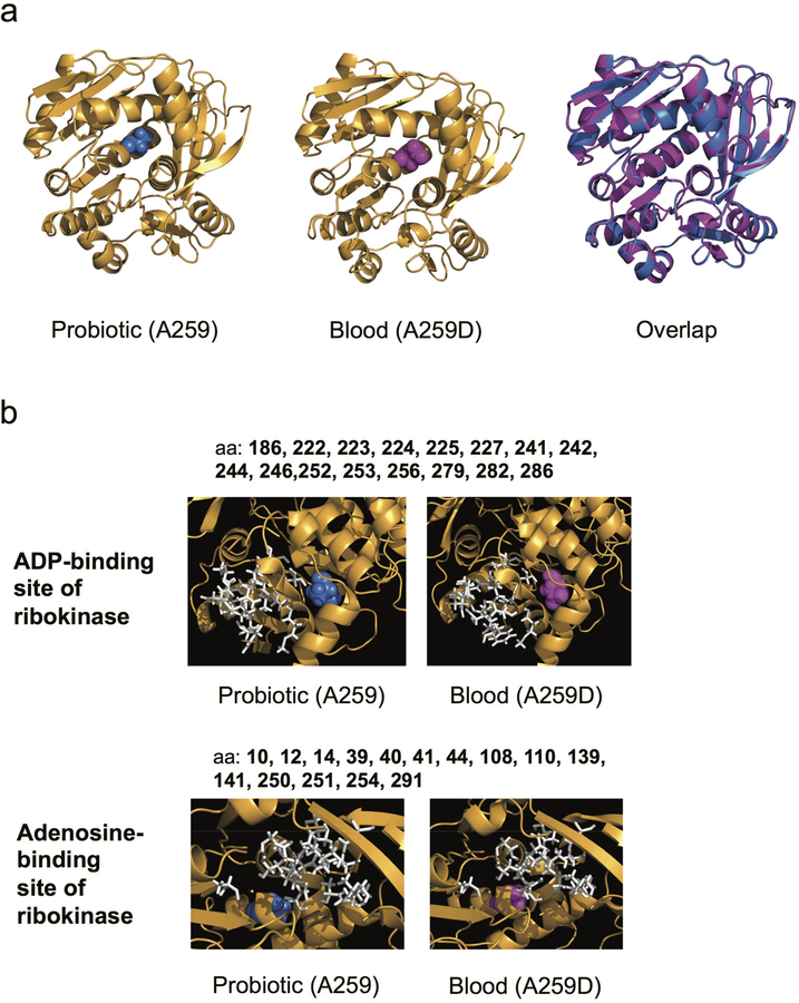
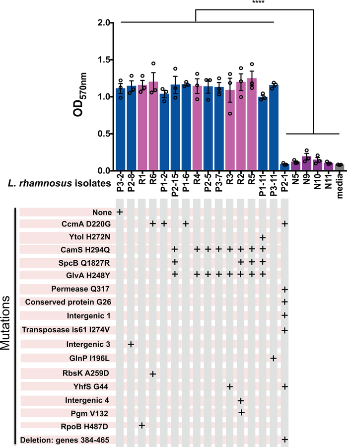
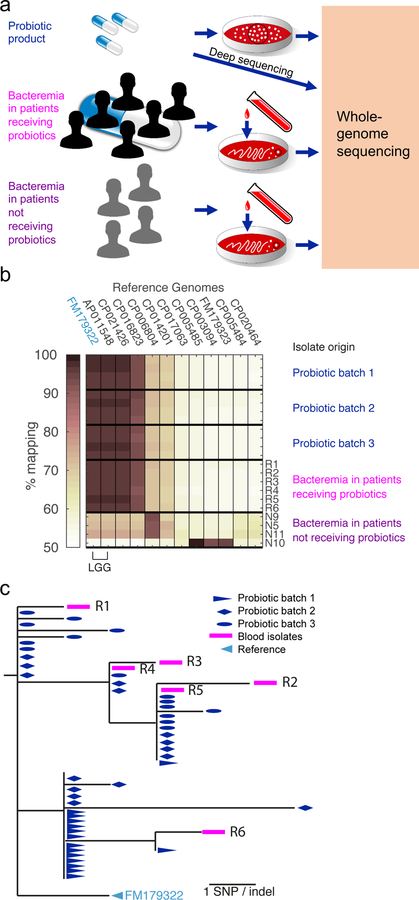
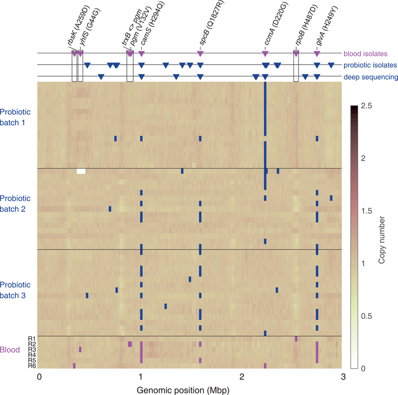
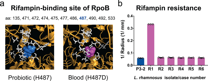
Probiotics are routinely administered to hospitalized patients for many potential indications1 but have been associated with adverse effects that may outweigh their potential benefits2-7. It is particularly alarming that probiotic strains can cause bacteremia8,9, yet direct evidence for an ancestral link between blood isolates and administered probiotics is lacking. Here we report a markedly higher risk of Lactobacillus bacteremia for intensive care unit (ICU) patients treated with probiotics compared to those not treated, and provide genomics data that support the idea of direct clonal transmission of probiotics to the bloodstream. Whole-genome-based phylogeny showed that Lactobacilli isolated from treated patients' blood were phylogenetically inseparable from Lactobacilli isolated from the associated probiotic product. Indeed, the minute genetic diversity among the blood isolates mostly mirrored pre-existing genetic heterogeneity found in the probiotic product. Some blood isolates also contained de novo mutations, including a non-synonymous SNP conferring antibiotic resistance in one patient. Our findings support that probiotic strains can directly cause bacteremia and adaptively evolve within ICU patients.


Conflict of interest statement

Competing Interests Statement

The authors have no competing interests as defined by Nature Research, or other interests that might be perceived to influence the interpretation of the article.

Figures

Extended Data Figure 1.
Deep sequencing identifies loci of diversity across probiotic product batches. Five probiotic batches (batches P2–P6, see Supplementary Table 2) were sequenced at high depth together with a single colony. In each batch, for each position in the reference genome, a two-sided Fisher exact test was carried out to determine differences in diversity between the batch derived sequences and the colony derived ones, and the respective p-values were plotted. Significant loci (p-value<1.66e-8) are marked with labels A-O (for details see Supplementary Table 6). A single locus of increased diversity in the colony in comparison to only one of the probiotic batches was also observed (green).
Extended Data Figure 2.
Extended Data Figure 2.. The blood-isolate-specific rpoB SNP does not perturb the RpoB predicted structure but occurs near the DNA-binding site and is associated with rifampin resistance in other bacterial species.
(a) Predicted structures of L. rhamnosus GG RNA polymerase β-subunit RpoB with histidine at position 487 seen in the probiotic (blue, left), aspartic acid at position 487 seen in the blood isolate from Patient R1 (magenta, middle), and overlap (right). (b) Predicted DNA-binding site amino acids are shown in white, with the histidine (blue) of the probiotic (left) and the aspartic acid (magenta) of blood isolate from Patient R1 (right) shown compared to the DNA-binding positions. (c) Amino acid (aa) sequence alignment of the Rifampin cluster I of the RpoB protein from L. rhamnosus GG and other genera. Numbering begins and ends at the first and last aa of the cluster; asterisks depict evolutionarily conserved aa residues; red asterisk shows the conservation across species of the histidine. In magenta, aa substitution H487D of the L. rhamnosus GG rifampin-resistant isolate (Patient R1) found in this study, H481D of S. aureus M1112 rifampin-resistant isolate, and H482D of B. velezensis rifampin-resistant isolate; in orange, substitution H481Y of S. epidermidis RP62A rifampin-resistant isolate, H489Y of E. faecium 343–3 rifampin-resistant isolate, H489Y of E. faecium 40–4 rifampin-resistant isolate, H526Y of E. coli K-12 substr. MG1655 rifampin-resistant isolate, and H482Y of B. velezensis rifampin-resistant isolate; in lavender, substitution H489Q of E. faecium 38–15 rifampin-resistant isolate; in brown, substitution H482R of B. velezensis rifampin-resistant isolate; in turquoise, substitution H482C of B. velezensis rifampin-resistant isolate.
Extended Data Figure 3.
Extended Data Figure 3.. The blood-isolate-specific ribokinase SNP does not perturb the predicted structure of ribokinase but occurs near the active site.
(a) Predicted structures of probiotic ribokinase with A259 (blue, left), blood isolate from Patient R1 with ribokinase A259D SNP (magenta, middle) and overlap (right). (b) The predicted binding site amino acids of ribokinase for adenosine are shown in white, with the alanine 259 (blue) of the probiotic (left) and the aspartic acid (magenta) of blood isolate 1 (right) shown compared to the adenosine-binding positions.
Extended Data Figure 4.
Extended Data Figure 4.. Biofilm formation of probiotic and blood L. rhamnosus isolates.
Blood isolates from patients receiving (R1–R6) and those not receiving probiotics (N5, N9, N10, N11), as well as selected probiotic isolates, were tested for biofilm formation. Isolates are grouped by similar mutations, as depicted in the grid below the isolate labels. Isogenic probiotic isolates from different probiotic capsules were used as controls, if available, as were controls for mutations found in blood isolates, when available. Px-y, were x = probiotic batch number, y = probiotic isolate number. Bars depict means of three independent experiments performed on different days, with 3 technical replicates per isolate in each experiment. Error bars depict the SEM. **** P<0.0001 by ANOVA test followed by Tukey’s multiple comparisons test for the pairwise comparison of any of the isolates making biofilm (defined as OD570 >1) compared to either P2–1, N5, N9, N10, N11, or medium control. There were no statistically significant differences among the isolates making biofilm or among the isolates not making biofilm.
Fig. 1.
Fig. 1.. Genomic evidence for Lactobacillus rhamnosus transmission from probiotic capsule to the blood of patients.
(a) Schematic for whole-genome sequencing of Lactobacillus rhamnosus probiotic isolates, blood isolates from ICU patients (n=6) receiving probiotics, and blood isolates from non-ICU patients (n=4) who were not receiving probiotics. Black circles represent sequencing multiple individual colonies for each probiotic batch but a single colony for each blood isolate. (b) Similarity between L. rhamnosus isolates and available reference genomes shown as the fraction of reads aligned to each reference. Isolates are identified by their source: four representative isolates from each of three probiotic product batches, the six blood isolates from patients receiving probiotics, and the four blood L. rhamnosus isolates from patients not receiving probiotics. (c) Phylogenetic analysis of all 54 sequenced L. rhamnosus GG (LGG) isolates: 16 isolates from each of 3 separate probiotic batches (blue), and the 6 blood isolates from Patients R1 to R6 (magenta).
Fig. 2.
Fig. 2.. Coverage of the LGG reference genome for the probiotic and blood Lactobacillus rhamnosus isolates.
For each isolate (row in matrix) SNPs are marked as squares (magenta for blood isolates, blue for product isolates). Triangles (top panel) indicate all mutations identified in blood isolates (magenta triangles) and probiotic product (blue triangles) compared to the LGG reference genome FM179322. For the probiotic product, these are either high-quality SNPs in whole-genome sequencing (middle row) or diversity identified by deep sequencing of the product (bottom row, see Methods). Annotation is included for all SNPs identified in blood isolates. SNPs identified only in blood isolates are indicated with black frame.
Fig 3.
Fig 3.. The Lactobacillus rhamnosus blood-isolate-specific rpoB SNP occurs at the rifampin-binding site and confers rifampin resistance.
(a) Predicted structure of L. rhamnosus GG RNA polymerase β-subunit RpoB showing the rifampin-binding site (white) with histidine 487 of the probiotic (blue, left) and aspartic acid 487 of the blood isolate from Patient R1 (magenta, right). (b) Rifampin susceptibility testing of blood isolates of each patient (R1–R6) compared to a probiotic isolate with no SNPs (P3–2). Bars depict the medians of 3 independent experiments, and error bars show the interquartile ranges. *P = 0.0021 for R1 compared to P3–2 by Kruskal- Wallis test followed by Dunn’s multiple comparisons test. The blood isolate from Patient R1 was resistant based on zone cutoffs for S. aureus (Supplementary Table 8).

Comment in

References

    1. Sarah HY, Jernigan JA & McDonald LC Prevalence of probiotic use among inpatients: a descriptive study of 145 US hospitals. Am. J. Infect. Control 44, 548–553 (2016). - PMC - PubMed
    1. Szajewska H What are the indications for using probiotics in children? Arch. Dis. Child 101, 398–403 (2016). - PubMed
    1. Barraud D, Bollaert P-E & Gibot S Impact of the administration of probiotics on mortality in critically ill adult patients: a meta-analysis of randomized controlled trials. Chest 143, 646–655 (2013). - PubMed
    1. Barraud D et al. Probiotics in the critically ill patient: a double blind, randomized, placebo-controlled trial. Intensive Care Med. 36, 1540–1547 (2010). - PubMed
    1. Honeycutt TCB et al. Probiotic administration and the incidence of nosocomial infection in pediatric intensive care: a randomized placebo-controlled trial. Pediatr. Crit. Care Med. 8, 452–8; quiz 464 (2007). - PubMed

Publication types

MeSH terms