Case Reports
. 2019 Oct;10(Suppl 1):S20-S25.
doi: 10.1016/j.jcot.2019.03.025. Epub 2019 Apr 1.

Allogenic umbilical cord blood-derived mesenchymal stem cells implantation for the treatment of juvenile osteochondritis dissecans of the knee

Affiliations
Case Reports

Allogenic umbilical cord blood-derived mesenchymal stem cells implantation for the treatment of juvenile osteochondritis dissecans of the knee

Jun-Seob Song et al. J Clin Orthop Trauma. 2019 Oct.

Abstract

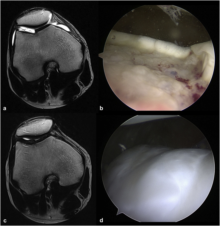
Background: Osteochondritis dissecans (OCD) is a pathologic condition accompanied by the gradual destruction of subchondral bone and defects in the overlying articular cartilage.This case series reports the results of allogenic human umbilical cord blood-derived mesenchymal stem cell (hUCB-MSC) implantation for the treatment of osteochondral defect in two cases of juvenile osteochondritis dissecans.

Case presentation: Two patients with osteochondral defect of the knee recovered from the disease enough to begin major exercise 1 year after hUCB-MSCs implantation. The IKDC, VAS, and Tegner score of the two patients showed an excellent improvement and concurrent arthroscopy was performed; cartilage regeneration of ICRS grade 1 similar to normal was observed. The modified two-dimensional MOCART scores increased in both cases over time.

Conclusion: This is the first case series detailing the results of treating juvenile OCD lesions using hUCB-MSCs. This could be an option for treating juvenile OCD.

Keywords: Mesenchymal stem cells; Osteochondral defect; Osteochondritis dissecans; Umbilical cord blood.


Figures

Fig. 1
Fig. 1
a) The left vial contains hyaluronic acid and right vial contains hUCB-MSCs. b) Aspirated hUCB-MSCs are injected into the hyaluronic acid vial. c) We mix the hUCB-MSCs with hyaluronic acid. d) The mixture of hUCB-MSCs and hyaluronic acid is put in a syringe. e) After making subchondral holes. f) After injection of the mixture into subchondral holes.
Fig. 2
Fig. 2
a. b) Pre-operative coronal and sagittal fat-suppressed proton-weighted MR images of left knee in a 16-year-old male show an osteochochondritis dissecans lesion at lateral femoral condyle, a linear high signal intensity line between the bone fragment and lateral femoral condyle as well as multiple subchondral cysts. c) An arthroscopic image demonstrates cartilage defect and exposed irregular subchondral bone surface of the lateral femoral condyle d. e) Post-operative coronal and sagittal proton-weighted MR images of the left knee at 33 months reveal articular cartilage regeneration (modified 2D MOCART score of 60). Subchondral cysts are still noted but the number and size of cysts are markedly decreased. f) A concurrent arthroscopic image at 35 months depicts cartilage regeneration at hUCB-MSCs implantation site on the lateral femoral condyle.
Fig. 3
Fig. 3
a) Axial T2-weighted MR image of the right knee in a 16-year-old male reveals a full thickness cartilage defect at the trochlea lateral side. b) An arthroscopic image demonstrates cartilage defect and an exposed irregular subchondral bone surface of the trochlea. c) Axial T2-weighted MR image demonstrates cartilage regeneration at the trochlea osteochondritis dissecans lesion after hUCB-MSCs implantation at 20 months. d ) A concurrent arthroscopic image at 20 months depicts cartilage regeneration after hUCB-MSCs implantation at the trochlea.
Fig. 4
Fig. 4
a) IKDC scores of case 1 and case 2. b) VAS scores of case 1 and case 2. c) Tegner scores of case 1 and case 2.

References

    1. Defierline A.J., Goldsfein J.L., Rue J.-P.H., Bach B.R., Jr. Evaluation and treatment of osteochondritis dissecans lesions of the knee. J Knee Surg. 2008;21:106–115. - PubMed
    1. Steinhagen J., Bruns J., Deuretzbacher G., Ruether W., Fuerst M., Niggemeyer O. Treatment of osteochondritis dissecans of the femoral condyle with autologous bone grafts and matrix-supported autologous chondrocytes. Int Orthop. 2010;34:819–825. - PMC - PubMed
    1. Pascual-Garrido C., Moran C.J., Green D.W., Cole B.J. Osteochondritis dissecans of the knee in children and adolescents. Curr Opin Pediatr. 2013;25:46–51. - PubMed
    1. Juneau C., Paine R., Chicas E., Gardner E., Bailey L., McDermott J. Current concepts in treatment of patellofemoral osteochondritis dissecans. Int J Sports Phys Ther. 2016;11:903. - PMC - PubMed
    1. Brittberg M., Lindahl A., Nilsson A., Ohlsson C., Isaksson O., Peterson L. Treatment of deep cartilage defects in the knee with autologous chondrocyte transplantation. N Engl J Med. 1994;331:889–895. - PubMed

Publication types