

A novel single myocapsular sleeve (SMS) repair technique to reduce dislocation in posterior approach to the hip: A clinico-radiographic study
- PMID: 31700214
- PMCID: PMC6823894
- DOI: 10.1016/j.jcot.2019.03.014
A novel single myocapsular sleeve (SMS) repair technique to reduce dislocation in posterior approach to the hip: A clinico-radiographic study
Erratum in
-
Erratum regarding previously published articles.J Clin Orthop Trauma. 2020 Nov-Dec;11(6):1169-1171. doi: 10.1016/j.jcot.2020.09.032. Epub 2020 Sep 26. J Clin Orthop Trauma. 2020. PMID: 33013141 Free PMC article.
-
Erratum regarding previously published articles.J Clin Orthop Trauma. 2021 Aug 5;21:101556. doi: 10.1016/j.jcot.2021.101556. eCollection 2021 Oct. J Clin Orthop Trauma. 2021. PMID: 34414070 Free PMC article.
Abstract
Background: To assess a new modification of posterior approach to the hip and its effect on stability and functional outcome in total hip arthroplasty.
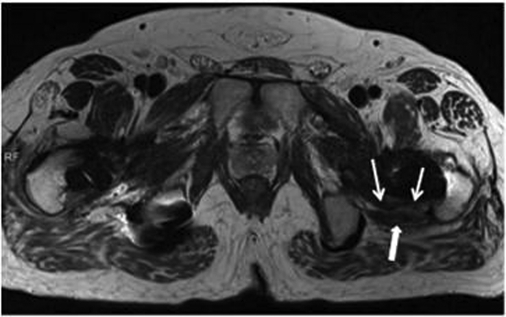
Material & methods: A comparative retrospective study was done to assess the functional outcome and rate of dislocation among 233 hips (Group A) operated by conventional posterior approach and 567 hips (Group B) by our novel modified posterior approach. In this technique, 2-3 stay sutures are applied in external rotators, then a single conjoint-myocapsular sleeve is raised linearly over the capsule with adherent fibers of gluteus minimus to piriformis tendon, short rotators and part of quadratus for exposure of femoral head. After inserting the definite prosthesis, upper part of sleeve (capsule, piriformis tendon) is sutured at the lower part of tip of greater trochanter & lower part with lateral trochanteric bone. Fifty patients, using randomised tables, in group B underwent MRI to evaluate the efficacy of the repair at 1 and 12 weeks postoperatively.
Results: Average Harris hip score at minimum 3.9 year follow up was 83.2 in Group A & 88.7 in Group B. Group B had only one dislocation (0.176%) while Group A had 12 dislocations (5.15%). MRI showed intact repair in 47 patients (94%); fibrous continuity in 2 patients (6%) in group B patients.
Conclusion: Intermediate results shows that this technique provides enhanced stability and improved functional outcome. But more prospective and randomised controlled studies with long term followup are required to confirm its role in prevention of hip dislocations.
Keywords: Dislocation; Hip arthroplasty; Posterior hip approach; SMS technique; Single myocapsular sleeve.
© 2019 Delhi Orthopedic Association. All rights reserved.
Figures


References
-
- Vasilakis I., Solomou E., Vitsas V., Fennema P., Korovessis P., Siamblis D.K. Correlative analysis of MRI-evident abductor hip muscle degeneration and power after minimally invasive versus conventional unilateral cementless THA. Orthopedics. Dec. 2012;35(12):e1684–e1691. - PubMed
-
- Gibson A. Posterior exposure of the hip joint. J Bone Joint Surg Br Vol. May 1950;32-B(2):183–186. - PubMed
-
- Fackler C.D., Poss R. Dislocation in total hip arthroplasties. Clin Orthop. Sep. 1980;151:169–178. - PubMed
-
- McCollum D.E., Gray W.J. Dislocation after total hip arthroplasty. Causes and prevention. Clin Orthop. Dec. 1990;261:159–170. - PubMed
LinkOut - more resources
Full Text Sources
