. 2019 Nov 4:15:64.
doi: 10.1186/s13223-019-0379-5. eCollection 2019.

Differential expression of CCR2 and CX3CR1 on CD16+ monocyte subsets is associated with asthma severity

Affiliations

Differential expression of CCR2 and CX3CR1 on CD16+ monocyte subsets is associated with asthma severity

Reem Al-Rashoudi et al. Allergy Asthma Clin Immunol. .

Abstract

Background: Monocytes play an important role in immune and inflammatory diseases and monocyte subsets are predictors of disease in certain conditions. Expression of the chemokine receptors, CCR2 and CX3CR1 on monocyte subsets relates to their function and can be used in their characterization. Our objective was to determine whether CD14, CD16, CCR2 and CX3CR1 on monocyte subsets are potential indicators of asthma severity.

Methods: Blood samples were collected from Saudi Arabian patients with asthma and normal healthy individuals. Six-color flow-cytometry phenotypic analysis was used to identify human blood monocyte subsets, based on their expression of CD14 and CD16 following CD45 gating. Expression of CCR2 and CX3CR1 was analysed on classical (CD14++CD16-), intermediate (CD14++CD16+) and non-classical (CD14+CD16++) subsets and correlated with disease severity.

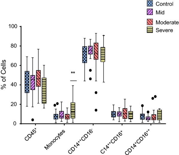
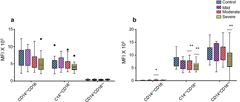
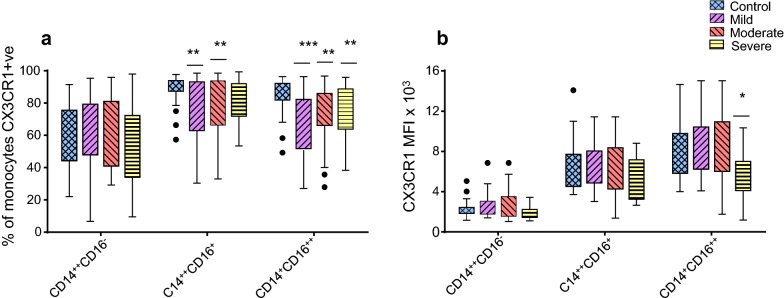
Results: We demonstrated a significant increase in percentage of total CD45-positive monocytes in the blood of patients with severe asthma, but the proportion of the individual monocyte subsets was not significantly changed when patients with mild, moderate and severe asthma were compared with healthy individuals. CD16 expression (mean fluorescence intensity, MFI) was decreased on intermediate and non-classical subsets in patients with severe asthma compared to healthy controls. CX3CR1 expression was also lower, with a lower percentage of cells expressing CX3CR1 in the non-classical CD14+CD16++ subset in all patients with asthma and this was inversely related to the percentage of cells expressing CCR2.

Conclusions: CCR2 expression on monocytes indicated a tendency toward more phagocytic monocytes in patients with asthma. The differential expression of CD16, CX3CR1 and CCR2 on monocyte subsets in peripheral blood indicates modulation of the inflammatory response and suggests a role for monocytes in asthma pathogenesis.

Keywords: Biomarker; CD14; Chemokine receptor; Flow cytometry; Saudi Arabia.


Conflict of interest statement

Competing interestsThe authors declare that they have no competing interests.

Figures

Fig. 1
Fig. 1
Gating strategy for defining monocyte subsets. a A side scatter (SSC) versus forward side scatter (FSC) dot plot was used to identify cells from debris (P1); b a SSC/CD45 dot plot from live gate (P2) was used to gate on CD45+ cells and exclude large granulocytes; c a FSC/SSC dot plot from the CD45+ expressing cells was used to tightly gate on the monocyte population (positioned just above the lymphocytes); d a CD14/CD16 dot plot from CD14+ monocytes was arbitrarily set based on their fluorescent intensity and on FMO and isotype controls to reveal the three monocyte subsets (CD14++CD16, CD14++CD16+, and CD14+CD16++); e the expression of CCR2 and CX3CR1 by the three monocyte subsets was set based on FMO and isotype control
Fig. 2
Fig. 2
Comparative analysis of the percentage of CD45+ cells, total monocyte population and monocyte subsets in mild asthmatic patients (n = 22), moderate asthmatic patients (n = 32) and severe asthmatic patients (n = 22) compared to healthy control patients (n = 35). Each monocyte subset is shown as box groups where the left box indicates healthy subjects and the next boxes indicate mild, moderate and severe asthmatic patients respectively. The box ranges from the 25th to 75th percentile, where the top end of the whiskers indicates the largest value less than the sum of 75th percentile plus 1.5×IQR (interquartile range) and any values greater than this are considered outliers (represented by dots) and the bottom end of the whiskers indicates the lowest value greater than the 25th percentile minus 1.5×IQR and any values less than this are considered outliers. The middle line represents the median. Significance was calculated using Holm–Sidak unpaired multiple comparison test and is indicated with **(p < 0.01)
Fig. 3
Fig. 3
Comparative analysis of CD14 expression (a) and CD16 expression (b) on monocyte subsets from mild asthmatic patients (n = 22), moderate asthmatic patients (n = 32) and severe asthmatic patients (n = 22) compared to healthy control (n = 35). Each monocyte subset is shown as box groups (as defined in Fig. 2) where the left box indicates healthy subjects and the next boxes indicate mild, moderate and severe asthmatic patients respectively. Gating was performed as in Fig. 1 and significance, compared to healthy controls, was calculated using Holm–Sidak unpaired multiple comparison test and is indicated with *(p < 0.05), **(p < 0.01)
Fig. 4
Fig. 4
Comparative analysis of the percentage of monocytes that are CCR2 positive (a) or CCR2 expression (MFI; b) on monocyte subsets from mild asthmatic patients (n = 22), moderate asthmatic patients (n = 32) and severe asthmatic patients (n = 22) compared to healthy control patients (n = 35). Each monocyte subset is shown as box groups (as defined in Fig. 22 where the left box indicates healthy subjects and the next boxes indicate mild, moderate and severe asthmatic patients respectively. Gating was performed as in Fig. 1 and significance, compared to healthy controls, was calculated using Holm–Sidak unpaired multiple comparison test and is indicated with **(p < 0.01), ***(p < 0.001)
Fig. 5
Fig. 5
Comparative analysis of the percentage of monocytes that are CX3CR1 positive (a) or CX3CR1 expression (MFI; b) on monocyte subsets from mild asthmatic patients (n = 22), moderate asthmatic patients (n = 32) and severe asthmatic patients (n = 22) compared to healthy control patients (n = 35). Each monocyte subset is shown as box groups (as defined in Fig. 2) where the left box indicates healthy subjects and the next boxes indicate mild, moderate and severe asthmatic patients respectively. Gating was performed as in Fig. 1 and significance, compared to healthy controls, was calculated using Holm–Sidak unpaired multiple comparison test and is indicated with *(p < 0.05), **(p < 0.01), ***(p < 0.001)
Fig. 6
Fig. 6
Correlations shown are between the percentage of monocytes that are CCR2 or CX3CR1 positive in the CD14++CD16+ monocyte subset from mild (a) moderate (b) and severe (c) asthmatic patients and in the CD14+CD16++ monocyte subset from mild (d) moderate (e) and severe (f) asthmatic patients using nonparametric Spearman rank correlation Rs

Similar articles

Cited by

References

    1. Maslan J, Mims JW. What is asthma? Pathophysiology, demographics, and health care costs. Otolaryngol Clin North Am. 2014;47:13–22. doi: 10.1016/j.otc.2013.09.010. - DOI - PubMed
    1. Anandan C, Nurmatov U, van Schayck O, Sheikh A. Is the prevalence of asthma declining? Systematic review of epidemiological studies. Allergy. 2010;65:152–167. doi: 10.1111/j.1398-9995.2009.02244.x. - DOI - PubMed
    1. Nakagome K, Nagata M. Pathogenesis of airway inflammation in bronchial asthma. Auris Nasus Larynx. 2011;38:555–563. doi: 10.1016/j.anl.2011.01.011. - DOI - PubMed
    1. Holgate ST. Pathophysiology of asthma: what has our current understanding taught us about new therapeutic approaches? J Allergy Clin Immunol. 2011;128(3):495–505. doi: 10.1016/j.jaci.2011.06.052. - DOI - PubMed
    1. Lloyd CM, Saglani S. T cells in asthma: influences of genetics, environment, and T-cell plasticity. J Allergy Clin Immunol. 2013;131(5):1267–1274. doi: 10.1016/j.jaci.2013.02.016. - DOI - PubMed