

Linked color imaging for the detection of early gastrointestinal neoplasms
- PMID: 31700545
- PMCID: PMC6826899
- DOI: 10.1177/1756284819885246
Linked color imaging for the detection of early gastrointestinal neoplasms
Abstract
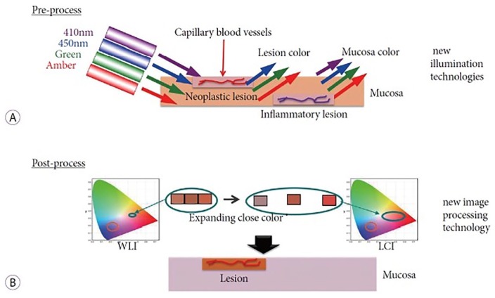
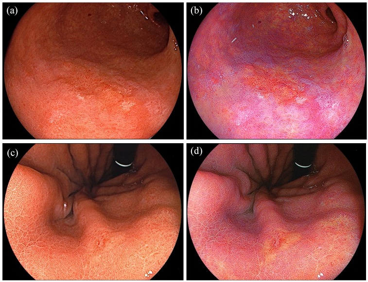
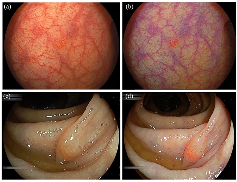
In routine upper and lower gastrointestinal endoscopy, overlooking neoplastic lesions is inevitable even for well-trained endoscopists. Various methods have been reported to improve the detection of gastrointestinal neoplasms including chromoendoscopy, special endoscopes, and processor and image enhanced technologies. Equipment-based image enhanced endoscopy (e-IEE) using narrow band imaging (NBI) and blue laser imaging (BLI) is useful to characterize known lesions with magnification at a close-up view. However, they are not useful for the early detection of superficial, pale neoplasms, or both because of the weak image at a distant view in a wide lumen such as the stomach or colon. Linked color imaging (LCI) is a novel pre- and post-processing technology developed by Fujifilm Corporation that has sufficient brightness to illuminate a wide lumen. LCI delineates early gastric cancers as orange-red and intestinal metaplasia as purple. LCI improves the adenoma detection rate in the colon and decreases the polyp miss rate. LCI contributes to the detection of superficial lesions throughout the gastrointestinal tract by enhancing the color contrast between the neoplasm and the surrounding mucosa. LCI can distinguish them by their specific color allocation based mainly on the distribution of capillaries. The authors believe that moving forward, LCI should be used in routine upper and lower gastrointestinal endoscopy.
Keywords: colon polyp; diagnosis; endoscopy; image enhanced endoscopy; linked color imaging.
© The Author(s), 2019.
Conflict of interest statement
Conflict of interest statement: H.Y. has a consultant relationship with Fujifilm Corporation and has received honoraria, grants, and royalties from the company. H.O. and Y.H. have received honoraria from Fujifilm Corporation. The other authors have no financial conflicts of interest.
Figures
Similar articles
-
Current status and future perspective of linked color imaging for gastric cancer screening: a literature review.J Gastroenterol. 2023 Jan;58(1):1-13. doi: 10.1007/s00535-022-01934-z. Epub 2022 Oct 26. J Gastroenterol. 2023. PMID: 36287268 Free PMC article. Review.
-
Recent Progress of Image-Enhanced Endoscopy for Upper Gastrointestinal Neoplasia and Associated Lesions.Dig Dis. 2024;42(2):186-198. doi: 10.1159/000535055. Epub 2023 Nov 10. Dig Dis. 2024. PMID: 37952532 Review.
-
Linked Color Imaging and Blue Laser Imaging for Upper Gastrointestinal Screening.Clin Endosc. 2018 Nov;51(6):513-526. doi: 10.5946/ce.2018.132. Epub 2018 Nov 2. Clin Endosc. 2018. PMID: 30384402 Free PMC article.
-
Appropriate Color Enhancement Settings for Blue Laser Imaging Facilitates the Diagnosis of Early Gastric Cancer with High Color Contrast.J Gastric Cancer. 2021 Jun;21(2):142-154. doi: 10.5230/jgc.2021.21.e13. Epub 2021 Jun 17. J Gastric Cancer. 2021. PMID: 34234976 Free PMC article.
-
Linked color imaging (LCI), a novel image-enhanced endoscopy technology, emphasizes the color of early gastric cancer.Endosc Int Open. 2017 Oct;5(10):E1005-E1013. doi: 10.1055/s-0043-117881. Epub 2017 Oct 10. Endosc Int Open. 2017. PMID: 29159276 Free PMC article.
Cited by
-
Rediscovering histology: what is new in endoscopy for inflammatory bowel disease?Therap Adv Gastroenterol. 2021 Apr 16;14:17562848211005692. doi: 10.1177/17562848211005692. eCollection 2021. Therap Adv Gastroenterol. 2021. PMID: 33948114 Free PMC article. Review.
-
Diagnostic ability of linked color imaging in ultraslim endoscopy to identify neoplastic lesions in the upper gastrointestinal tract.Endosc Int Open. 2022 Jan 14;10(1):E88-E95. doi: 10.1055/a-1723-2635. eCollection 2022 Jan. Endosc Int Open. 2022. PMID: 35047338 Free PMC article.
-
Advanced Imaging in Gastrointestinal Endoscopy: A Literature Review of the Current State of the Art.GE Port J Gastroenterol. 2022 Nov 4;30(3):175-191. doi: 10.1159/000527083. eCollection 2023 Jun. GE Port J Gastroenterol. 2022. PMID: 37387720 Free PMC article. Review.
-
Histopathological features of glandular atrophy of the lamina propria of the gastric mucosa during its occurrence and development.BMC Gastroenterol. 2023 Nov 15;23(1):395. doi: 10.1186/s12876-023-03033-6. BMC Gastroenterol. 2023. PMID: 37968594 Free PMC article.
-
Identification of gastric cancer with convolutional neural networks: a systematic review.Multimed Tools Appl. 2022;81(8):11717-11736. doi: 10.1007/s11042-022-12258-8. Epub 2022 Feb 18. Multimed Tools Appl. 2022. PMID: 35221775 Free PMC article.
References
-
- Kaltenbach T, Sano Y, Friedland S, et al. American gastroenterological association (AGA) institute technology assessment on image enhanced endoscopy. Gastroenterology 2008; 134: 327–340. - PubMed
-
- Dohi O, Yagi N, Naito Y, et al. Blue laser imaging-bright improves the real-time detection rate of early gastric cancer: a randomized controlled study. Gastrointest Endosc 2019; 89: 47–57. - PubMed
-
- Fukuda H, Miura Y, Osawa H, et al. Linked color imaging can enhance recognition of early gastric cancer by high color contrast to surrounding gastric intestinal metaplasia. J Gastroenterol 2019; 54: 396–406. - PubMed
Publication types
LinkOut - more resources
Full Text Sources
