. 2019 Nov 1;13(6):064101.
doi: 10.1063/1.5124827. eCollection 2019 Nov.

Rapid and multi-cycle smFISH enabled by microfluidic ion concentration polarization for in-situ profiling of tissue-specific gene expression in whole C. elegans

Affiliations

Rapid and multi-cycle smFISH enabled by microfluidic ion concentration polarization for in-situ profiling of tissue-specific gene expression in whole C. elegans

Gongchen Sun et al. Biomicrofluidics. .

Abstract

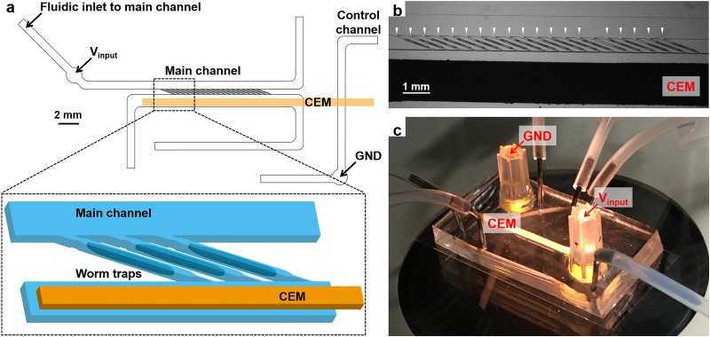
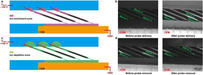
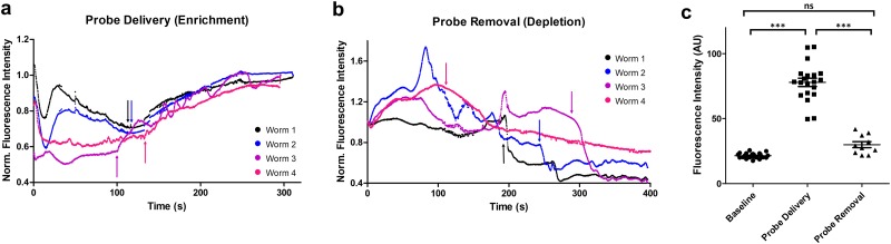
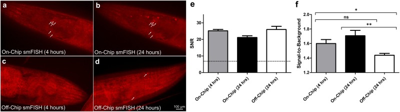
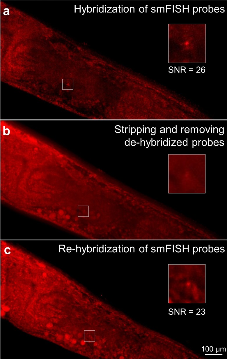
Understanding gene regulation networks in multicellular organisms is crucial to decipher many complex physiological processes ranging from development to aging. One technique to characterize gene expression with tissue-specificity in whole organisms is single-molecule fluorescence in situ hybridization (smFISH). However, this protocol requires lengthy incubation times, and it is challenging to achieve multiplexed smFISH in a whole organism. Multiplexing techniques can yield transcriptome-level information, but they require sequential probing of different genes. The inefficient macromolecule exchange through diffusion-dominant transport across dense tissues is the major bottleneck. In this work, we address this challenge by developing a microfluidic/electrokinetic hybrid platform to enable multicycle smFISH in an intact model organism, Caenorhabditis elegans. We integrate an ion concentration polarization based ion pump with a microfluidic array to rapidly deliver and remove gene-specific probes and stripping reagents on demand in individual animals. Using our platform, we can achieve rapid smFISH, an order of magnitude faster than traditional smFISH protocols. We also demonstrate the capability to perform multicycle smFISH on the same individual samples, which is impossible to do off-chip. Our method hence provides a powerful tool to study individual-specific, spatially resolvable, and large-scale gene expression in whole organisms.


Figures

FIG. 1.
FIG. 1.
(a) Schematic of the microfluidic/electrokinetic hybrid device. (b) Micrograph of individually loaded worms in worm traps. White arrowheads indicate individually loaded adult worms. (c) Image of the device setup.
FIG. 2.
FIG. 2.
(a) Operating principle of probe delivery. (b) Fluorescent images of FITC-Dextran (model of smFISH probes) transport into individual worms before and after the probe delivery process. The input reservoir received −150 V for 5 min. (c) Operating principle of probe removal. (d) Fluorescent images of FITC-Dextran transport out of individual worms before and after the probe removal process. The input reservoir received +150 V for 7 min. The image settings (brightness and contrast) are consistent for all fluorescent images in (b) and (d).
FIG. 3.
FIG. 3.
(a) Quantification of fluorescence intensity dynamics in individual worms during the probe delivery process. −150 V was applied at the input reservoir. Arrows indicate when the ion enrichment zone started to establish for each worm. (b) Quantification of fluorescence intensity dynamics in individual worms during the probe removal process. +150 V was applied at the input reservoir. Arrows indicate when the ion depletion front passed through each worm. (c) Quantification of fluorescence intensity of individual worms at baseline and after probe delivery and removal. A one-way ANOVA and Tukey's multiple comparison tests were performed. (***P < 0.001; ns, not significant).
FIG. 4.
FIG. 4.
Fluorescent images of individual puncta, each corresponding to a gpa-3 mRNA molecule in the neuronal cells. (a) and (b) On-chip probe delivery by the ICP-based ion pump in the same worm after (a) 4 h and (b) 24 h. (c) and (d) Off-chip diffusion-dominated delivery. (c) Individual puncta were not resolved after 4 h of off-chip incubation. (d) Puncta are visible only after 24 h of off-chip incubation. White arrows are added to aid puncta visualization. (e) Signal-to-noise ratio (SNR) of the puncta at each condition. The dotted line corresponds to the minimum value acceptable (SNR = 7). (f) Signal-to-background ratio of each punctum at each condition. An unpaired, two-tailed T test was performed (**P < 0.01, *P < 0.05; ns, not significant).
FIG. 5.
FIG. 5.
Multicycle smFISH of gpa-3 within the same worm using ICP-enhanced smFISH, and puncta were identified using FISH-quant. (a) Individual puncta were resolved after initial hybridization. (b) After DNase I treatment for 4 h, probes were dehybridized and removed by the ICP-based ion pump. (c) The same puncta were resolved again after rehybridization enhanced by ICP. Insets show the magnified area at the vicinity of the target mRNA molecule.

Similar articles

Cited by

References

    1. Schmid M., Davison T. S., Henz S. R., Pape U. J., Demar M., Vingron M., Scholkopf B., Weigel D., and Lohmann J. U., Nat. Genet. 37(5), 501–506 (2005). 10.1038/ng1543 - DOI - PubMed
    1. Tomancak P., Beaton A., Weiszmann R., Kwan E., Shu S., Lewis S. E., Richards S., Ashburner M., Hartenstein V., Celniker S. E., and Rubin G. M., Genome Biol. 3(12), research0088 (2002). 10.1186/gb-2002-3-12-research0088 - DOI - PMC - PubMed
    1. Lund J., Tedesco P., Duke K., Wang J., Kim S. K., and Johnson T. E., Curr. Biol. 12(18), 1566–1573 (2002). 10.1016/S0960-9822(02)01146-6 - DOI - PubMed
    1. Guarente L. and Kenyon C., Nature 408(6809), 255–262 (2000). 10.1038/35041700 - DOI - PubMed
    1. Elowitz M. B., Levine A. J., Siggia E. D., and Swain P. S., Science 297(5584), 1183–1186 (2002). 10.1126/science.1070919 - DOI - PubMed

LinkOut - more resources