

Lateral Extra-articular Tenodesis With Proximal Staple Fixation
- PMID: 31700777
- PMCID: PMC6823835
- DOI: 10.1016/j.eats.2019.03.020
Lateral Extra-articular Tenodesis With Proximal Staple Fixation
Abstract
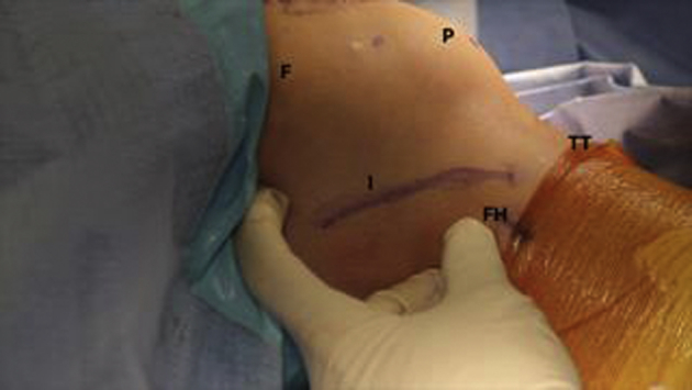
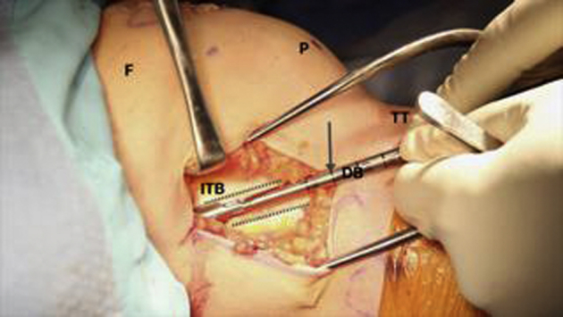
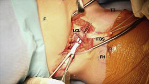
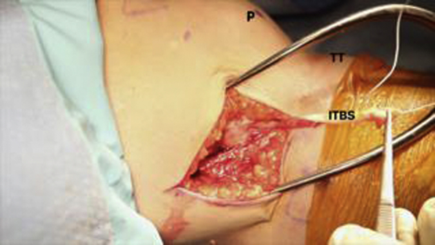
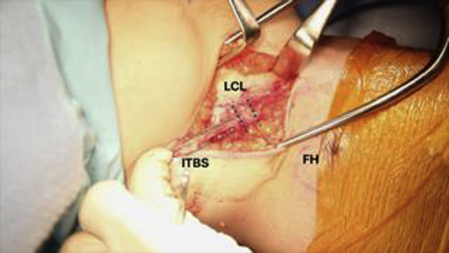
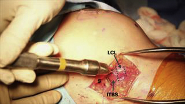
A technique for lateral extra-articular tenodesis using proximal staple fixation is described as an adjunct to anterior cruciate ligament (ACL) reconstruction. Lateral extra-articular tenodesis has been used in an effort to decrease failure rates in ACL-deficient patients with ligamentous laxity, prior failed ACL surgery, or grade 3 pivot-shift findings. Numerous surgeons have described combining ACL reconstruction with extra-articular surgery. The approach described in this article is easy and cost-effective. Moreover, because this technique uses a staple instead of a SwiveLock (Arthrex, Naples, FL) or other suture anchor, it limits the chance of conflicting with the tunnels for the ACL because there is no need to drill or punch additional tunnels.
© 2019 by the Arthroscopy Association of North America. Published by Elsevier.
Figures

References
-
- Galway H.R., MacIntosh D.L. The lateral pivot shift: A symptom and sign of anterior cruciate ligament insufficiency. Clin Orthop Relat Res. 1980;147:45–50. - PubMed
-
- Segond P. Aux Bureaux du Progrès Médical; Paris: 1879. Recherces cliniques et expérimentales sur les épanchements sanguins du genou par entorse. [in French]
-
- Buscayret C., Buscayret F., Farenq C. Intra- and extra-articular hamstring reconstruction of anterior cruciate ligament tears. Rev Chir Orthop Reparatrice Appar Mot. 2001;87:276–280. [in French] - PubMed
-
- Kennedy J., Jackson M.P., O’Kelly P., Moran R. Timing of reconstruction of the anterior cruciate ligament in athletes and the incidence of secondary pathology within the knee. J Bone Joint Surg Br. 2010;92:362–366. - PubMed
-
- Wroble R.R., Grood E.S., Cummings J.S., Henderson J.M., Noyes F.R. The role of the lateral extraarticular restraints in the anterior cruciate ligament-deficient knee. Am J Sports Med. 1993;21:257–262. discussion 263. - PubMed
LinkOut - more resources
Full Text Sources
