Review
. 2019 Jun 26;6(2):70-76.
doi: 10.1159/000495475. eCollection 2019 Apr-Jun.

South African Tick Bite Fever: An Overview

Affiliations
Review

South African Tick Bite Fever: An Overview

John Frean et al. Dermatopathology (Basel). .

Abstract

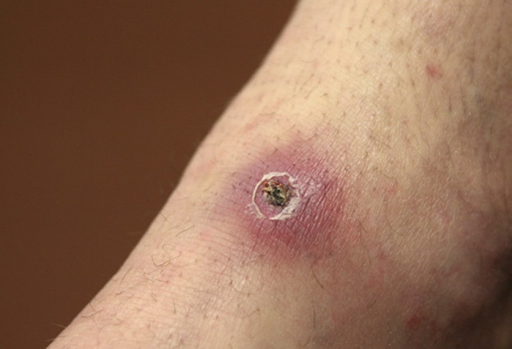
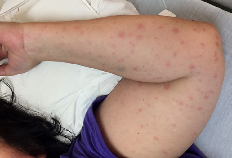
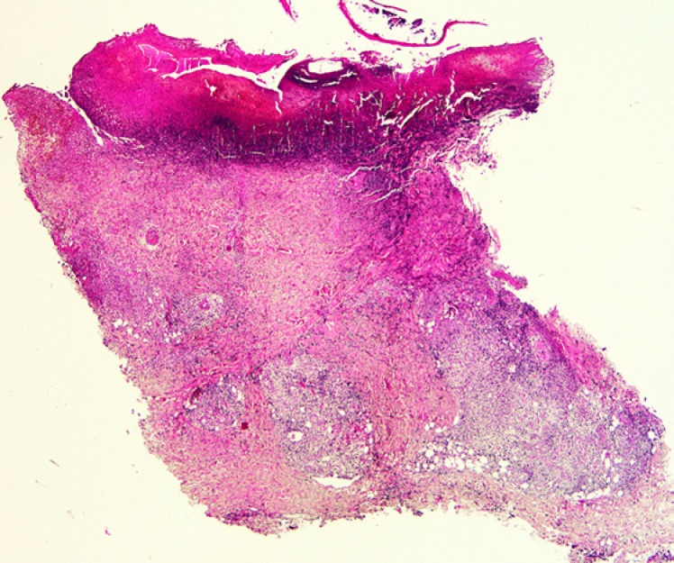
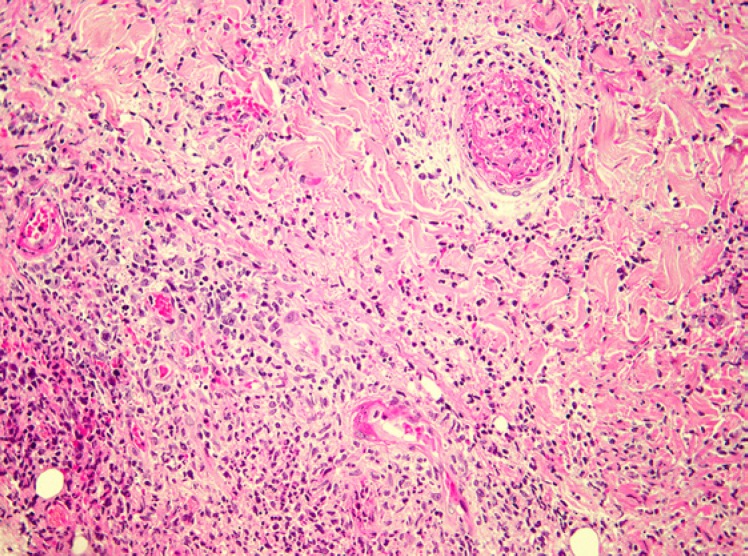
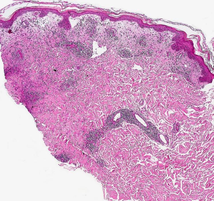
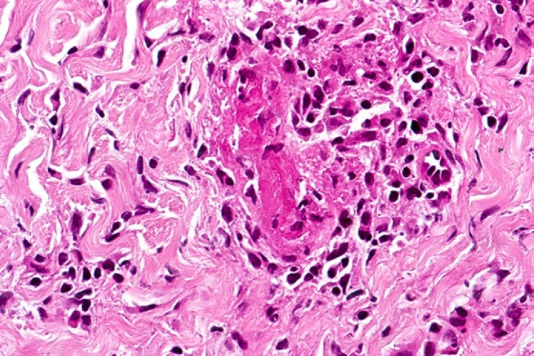
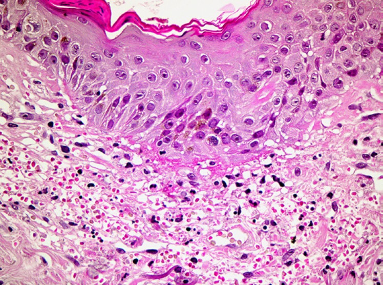
The rickettsiae are a diverse group of vector-borne zoonotic bacterial pathogens. The two common spotted fever diseases in existence in southern Africa are boutonneuse fever-like tick bite fever (TBF), caused by Rickettsia conorii, and African TBF, caused by R. africae. This review addresses demographic, epidemiological, clinical, diagnostic, therapeutic, and preventive aspects of TBF in the southern African context, including a discussion of the dermatopathological findings and potential diagnostic pitfalls.

Keywords: Rickettsia africae; Rickettsia conorii; South African tick bite fever.


Conflict of interest statement

The authors have no conflicts of interest to declare.

Figures

Fig. 1
Fig. 1
Clinical image of a typical eschar at the site of a tick bite.
Fig. 2
Fig. 2
Clinical image of a maculopapular skin rash associated with Rickettsia conorii infection.
Fig. 3
Fig. 3
Low-power view of a tick bite eschar. Note the inflammatory exudate overlying a zone of epidermal and superficial dermal necrosis, with a surrounding vital response.
Fig. 4
Fig. 4
Medium-power view of a tick bite eschar. Vasculitis and vascular thrombosis are evident below the base of the necrotic lesion, along with a background lymphohistiocytic and neutrophilic inflammatory infiltrate.
Fig. 5
Fig. 5
South African tick bite fever due to R. conorii infection. This low-power view shows a superficial and deep perivascular dermal lymphocytic infiltrate.
Fig. 6
Fig. 6
South African tick bite fever due to R. conorii infection. A lymphocytic vasculopathic reaction is observed in this high-power photomicrograph.
Fig. 7
Fig. 7
High-power photomicrograph of a skin biopsy from a proven case of South African tick bite fever due to R. conorii. There is conspicuous perivascular erythrocytic extravasation in the upper dermis in this example.

References

    1. Kelly P, Matthewman L, Beati L, Raoult D, Mason P, Dreary M, et al. African tick-bite fever: a new spotted fever group rickettsiosis under an old name. Lancet. 1992 Oct;340((8825)):982–3. - PubMed
    1. Pretorius AM, Birtles RJ. Rickettsia mongolotimonae infection in South Africa. Emerg Infect Dis. 2004 Jan;10((1)):125–6. 10.3201/eid0603.000309. - PMC - PubMed
    1. Pretorius AM, Birtles RJ. Rickettsia aeschlimannii: A new pathogenic spotted fever group rickettsia, South Africa. Emerg Infect Dis. 2002 Aug;8((8)):874. - PMC - PubMed
    1. Jensenius M, Fournier PE, Kelly P, Myrvang B, Raoult D. African tick bite fever. Lancet Infect Dis. 2003 Sep;3((9)):557–64. - PubMed
    1. Cohen GL, Blumberg LS, Karstaedt AS. Tick bite fever in black South Africans—a rare disease? J Infect. 1996 May;32((3)):235–7. - PubMed