. 2019 Sep;7(18):455.
doi: 10.21037/atm.2019.08.68.

microRNA-1 inhibits cardiomyocyte proliferation in mouse neonatal hearts by repressing CCND1 expression

Affiliations

microRNA-1 inhibits cardiomyocyte proliferation in mouse neonatal hearts by repressing CCND1 expression

Jingyi Gan et al. Ann Transl Med. 2019 Sep.

Abstract

Background: The functions of microRNA-1 (miR-1) in cardiac hypertrophy, and cardiomyocyte differentiation have been investigated. However, the mechanism on how miR-1 could repress cardiomyocyte proliferation has not been fully elucidated.

Methods: We address this issue by investigating whether miR-1 affected the proliferation of neonatal cardiomyocyte and identify some of the genes targeted by miR-1. miR-1 was over-expressed in neonatal cardiomyocytes and the effect on cell cycle and growth were analyzed by flow cytometry and Brdu-incorporation assay. Relevant vectors carrying the luciferase reporter were constructed for validation of miR-1 binding to its matching sites on the 3'-untranslated region of the predicated target mRNAs. Cardiomyocytes were co-transfected with the vectors and miR-1 mimics, then luciferase reporter assay was performed. Lastly, we examined the expression of target genes in cardiomyocytes after transfection with miR-1 mimics, as well as their normal expression pattern in 2- and 13-day-old mice hearts.

Results: We have demonstrated that miR-1 was the most significantly upregulated miRNA in 13-day-old mouse hearts compared with 2-day-old hearts. We also showed that miR-1 could repress cardiomyocyte G1/S phase transition, proliferation and viability. IGF1 and CCND1 were identified as candidate target genes regulated by miR-1. In addition, overexpression of miR-1 could suppress the expression of these two genes at the mRNA level. It could also correspondingly inhibit CCND1 expression at the protein level but not for IGF1.

Conclusions: Our results suggest that miR-1 plays an important role in inhibiting cardiomyocyte proliferation in the developing neonatal mouse heart by directly suppressing the cell-cycle regulator, CCND1.

Keywords: CCND1; cardiomyocyte proliferation; microRNA-1 (miR-1); neonatal mouse heart.


Conflict of interest statement

Conflicts of Interest: The authors have no conflicts of interest to declare.

Figures

Figure 1
Figure 1
Cardiomyocyte proliferation in 2- and 13-day-old mouse hearts. Immunofluorescent staining showing (A) the presence of proliferating (Ki-67+) cardiomyocytes (cTnT+) at day 2 and day 13 (yellow arrows). Bar charts showing (B) the percentage of cardiomyocytes that were Ki-67+ in 2- and 13-day-old hearts. Immunofluorescent staining showing (C) the presence of mitotic (pH3+) cardiomyocytes (cTnT+) at day 2 and day 13 (yellow arrows). Bar charts showing (D) the percentage of cardiomyocytes that were pH3+ in 2- and 13-day-old hearts. Values are presented as mean ± SD. scale bar: 50 µm. **, P<0.01.
Figure 2
Figure 2
microRNAs differentially expressed in 2- and 13-day-old mouse hearts. (A) Six significant differentially expressed miRNAs were identified in 2 and 13-day-old mouse hearts (performed in tri-plicate, P<0.05). (B) Stem Loop RT-qPCR analysis validated the miRNA microarray data that five miRNAs differentially expressed. Expression values are presented as mean ± SD. **, P<0.01. ns, no significance.
Figure 3
Figure 3
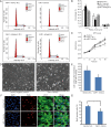
miR-1 inhibit cell cycle, proliferation and viability of neonatal cardiomyocytes. Cell cycle distribution (A) of cardiomyocytes after transfection with miR-1 mimics, mimics negative control (N.C), miR-1 antagomir and antagomir negative control. The cells transfected with miR-1 mimics showed an increased in G0/G1 phase and a decrease in S-phase compare with the mimics control (B). The height of the columns represents the viability of cardiomyocytes as determined by MTT assay (C). The cell density and number were determined under bright fields of microscope (D and E). Scale bar =20 µm. Cell proliferation was analyzed by BrdU-incorporation assay (F, scale bar =50 µm). Quantification of BrdU+ cardiomyocytes is presented as the percentage of BrdU+ cells out of the total number of DAPI+ cells (G). The values are presented as mean ± SD from three experiments. *, P<0.05; **, P<0.01. ns, no significance.
Figure 4
Figure 4
RT-qPCR assays. Validation of target genes predicated to be regulated by miR-1 in neonatal cardiomyocytes after transfection with miR-1 mimics (A), and also in 2- and 13-day-old hearts (B). Relative expression values are presented as mean ± SD. *P<0.05, **P<0.01. ns, no significance.
Figure 5
Figure 5
Ability of miR-1 to target the 3'UTR of IGF1 and CCND1 mRNA in neonatal cardiomyocytes. (A) Alignment of 5’ seeding region within miR-1 mature sequence and the putative binding sites within the 3'UTR region of IGF1, CCND1, Frs2 and FoxP1-mRNAs using TargetScan database. (B) Construct luciferase reporter containing the predicted binding sites within the 3'UTR of IGF1, CCND1, Frs2 and FoxP1 mRNAs into downstream of the firefly luciferase gene of pmirGLO vector. (C) Dual-Glo Luciferase reporter assays revealed reduced luciferase activity after co-transfection of pmirGLO-IGF1 3'UTR vector and pmirGLO-CCND1 3'UTR vector, respectively, with miR-1 mimics. The results were compared with luciferase activities from cardiomyocytes transfected with miR-1 mimics N.C. and pmirGLO empty vectors. The data are presented as mean ± SD from triplicated experiments. **, P<0.01 vs. miR-1 mimics NC or pmirGLO empty vectors.
Figure 6
Figure 6
Over-expression of miR1 decreases CCND1, but not IGF1, protein expression. Immunocytofluorescent staining revealed that CCND1 expression was significantly inhibited in cardiomyocytes following miR-1 mimics transfection (A and B). In contrast, no significant different difference in IGF1 expression in cTnT+ neonatal cardiomyocytes transfected with miR-1 mimics (C). Western blotting also confirmed that CCND1 expression was significantly down-regulated in cardiomyocytes after transfection with miR-1 mimics compared to the negative control (D). *, P<0.05. Scale bar =50 µm.
Figure 7
Figure 7
IGF-1 and CCND1 expression in 2- and 13-day-old hearts. The normal expression patterns of IGF1 and CCND1 were investigated in 2- and 13-day-old mouse hearts. Immunocytofluorescent staining revealed that CCND1 expression is down-regulated in 13-day-old hearts (A and B). In contrast, no significant change in IGF1 expression between 2- and 13-day-old mouse hearts (C). Values are presented as mean ± SD. scale bar =50 µm. **, P<0.01. ns, no significance.

References

    1. Huang Y, Harrison MR, Osorio A, et al. Igf Signaling is Required for Cardiomyocyte Proliferation during Zebrafish Heart Development and Regeneration. PLoS One 2013;8:e67266. 10.1371/journal.pone.0067266 - DOI - PMC - PubMed
    1. Zhang R, Han P, Yang H, et al. In vivo cardiac reprogramming contributes to zebrafish heart regeneration. Nature 2013;498:497-501. 10.1038/nature12322 - DOI - PMC - PubMed
    1. Tang MK, Kindler PM, Cai DQ, et al. Heart-type fatty acid binding proteins are upregulated during terminal differentiation of mouse cardiomyocytes, as revealed by proteomic analysis. Cell Tissue Res 2004;316:339-47. 10.1007/s00441-004-0881-y - DOI - PubMed
    1. Liu Y, Tang MK, Cai DQ, et al. Cyclin I and p53 are differentially expressed during the terminal differentiation of the postnatal mouse heart. Proteomics 2007;7:23-32. 10.1002/pmic.200600456 - DOI - PubMed
    1. Porrello ER, Mahmoud AI, Simpson E, et al. Transient Regenerative Potential of the Neonatal Mouse Heart. Science 2011;331:1078-80. 10.1126/science.1200708 - DOI - PMC - PubMed