Review
. 2020 Aug;11(4):553-579.
doi: 10.1007/s12975-019-00744-5. Epub 2019 Nov 7.

Neurovascular Unit as a Source of Ischemic Stroke Biomarkers-Limitations of Experimental Studies and Perspectives for Clinical Application

Affiliations
Review

Neurovascular Unit as a Source of Ischemic Stroke Biomarkers-Limitations of Experimental Studies and Perspectives for Clinical Application

Aleksandra Steliga et al. Transl Stroke Res. 2020 Aug.

Abstract

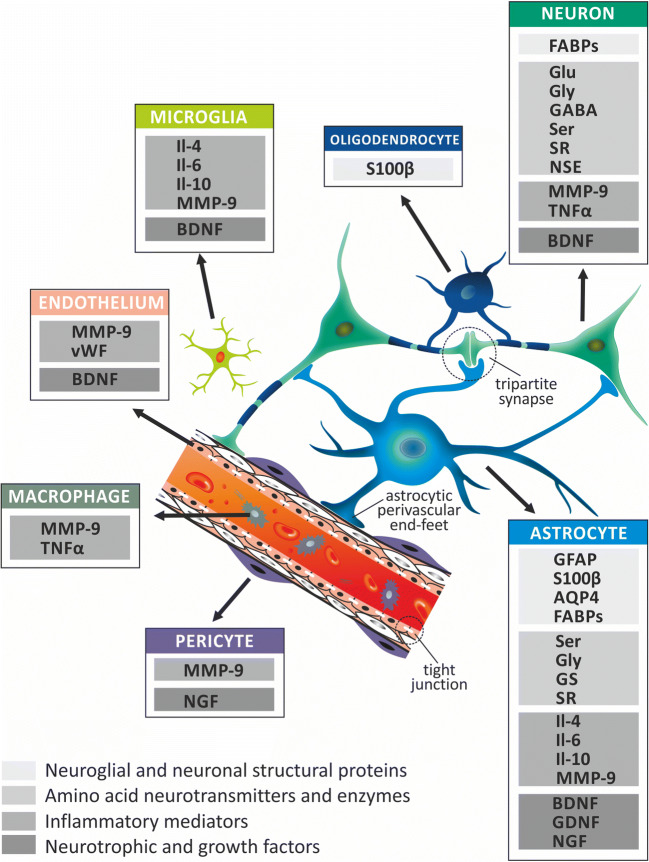
Cerebral stroke, which is one of the most frequent causes of mortality and leading cause of disability in developed countries, often leads to devastating and irreversible brain damage. Neurological and neuroradiological diagnosis of stroke, especially in its acute phase, is frequently uncertain or inconclusive. This results in difficulties in identification of patients with poor prognosis or being at high risk for complications. It also makes difficult identification of these stroke patients who could benefit from more aggressive therapies. In contrary to the cardiovascular disease, no single biomarker is available for the ischemic stroke, addressing the abovementioned issues. This justifies the need for identifying of effective diagnostic measures characterized by high specificity and sensitivity. One of the promising avenues in this area is studies on the panels of biomarkers characteristic for processes which occur in different types and phases of ischemic stroke and represent all morphological constituents of the brains' neurovascular unit (NVU). In this review, we present the current state of knowledge concerning already-used or potentially applicable biomarkers of the ischemic stroke. We also discuss the perspectives for identification of biomarkers representative for different types and phases of the ischemic stroke, as well as for different constituents of NVU, which concentration levels correlate with extent of brain damage and patients' neurological status. Finally, a critical analysis of perspectives on further improvement of the ischemic stroke diagnosis is presented.

Keywords: Astrocytes; Biomarkers; Neuroglia; Neurovascular unit; Stroke.


Conflict of interest statement

The authors declare that they have no conflict of interest.

Figures

Fig. 1
Fig. 1
Summary of pathophysiological processes developing in the course of ischemic stroke. The consequences of ischemic stroke result from processes beginning in a strictly defined time sequence. A decrease in cerebral blood flow results in reduction of oxygen and glucose delivered to the brain tissue. This initiates a cascade of biochemical processes ultimately leading to cellular destruction and death. One of the earliest consequences of these processes is change of oxidative glycolysis into less effective anaerobic pathway, followed by decrease in ATP production and raising lactate concentration. This results in decrease in all energy-dependent metabolic processes and dysfunction of the ion pumps leading to changes of ion concentrations (i.e., decrease in K+ intracellular levels and increase in Cl, Na+, and Ca2+ levels). This in turn contributes to influx of water and development of brain swelling. Depolarization of neuronal cell membranes leads to release of excessive amounts of glutamate and triggering glutamatergic excitotoxicity. Stimulating effect of glutamate, by changing Ca2+ concentration and activation of enzymes (e.g., proteases, lipases, phosphatases, and endonucleases), leads to further cell destruction. This is accompanied by the activation of oxidative stress along with an increase in production of free oxygen and nitrogen radicals. A further consequence of these processes is triggering of inflammatory response and release of proinflammatory cytokines and chemokines which destructive action affects all elements of neurovascular unit (NVU). In cerebral vessels, increase in permeability of BBB and damage to the endothelial cells occur, accompanied by up-regulation of thrombotic mechanisms. These processes are accompanied by apoptotic and necrotic cell death, which are dependent on the length of cerebral blood flow reduction, extent of the energetic metabolism disturbances, and localization of the cells (within infarct core or penumbra). Decrease in the cerebral blood flow and reduced availability of oxygen and glucose initiate the acute phase (up to 24 h from ischemia onset) processes in the course of ischemic stroke, i.e., glutamatergic excitotoxicity, increase in Ca2+ levels, and anaerobic glycolysis, leading to reduced efficiency of the energetic metabolism. The subacute phase (up to 7 days from ischemia onset) is characterized by occurrence of various forms of cell death, the blood–brain barrier (BBB) disintegration and leakage, as well as, initiation of the inflammatory response followed by release of mediators exacerbating the effects of primary damage. In contrast to the earlier phases, during the late phase (starting one week from ischemia onset), the initiated processes lead to limiting of deleterious effects of the cerebral blood flow reduction through development of reactive gliosis and gliotic scar, which enables demarcation of the necrotic infarct core from the surrounding intact tissue. At this phase, the reparative processes resulting from cell proliferation and differentiation dominate, what is reflected in intensive reconstruction of the cellular populations, angiogenesis and re-myelination.
Fig. 2
Fig. 2
The neurovascular unit (NVU) as a source of ischemic stroke biomarkers. The NVU concept has been proved useful for analysis of spatial and functional relationships among constituents of brain tissue. An important role of NVU is attributed to its characteristic morphological structures, such as tripartite synapses, astrocytic perivascular end-feet and vascular tight junctions. In accordance with the NVU concept, ischemic stroke biomarkers can be categorized as the representatives of either individual cell type or its several components. In addition, this concept enables division of ischemic stroke biomarkers into groups characterized by similar structure and functions representing the following categories: (1) neuroglial and neuronal structural proteins, (2) amino acid neurotransmitters and enzymes, (3) inflammatory mediators, and (4) neurotrophic and growth factors. While planning the research on the new ischemic stroke biomarkers, it is important to take into account that various components of NVU play different roles in the ischemic metabolic processes (e.g., oxidative and anaerobic glycolysis); signaling pathways (e.g., glutamate-glutamine shuttle, Ca2+ ion- and purines-based signaling) reveal different sensitivity to decreased cerebral blood perfusion, as well as, reveal different proliferation potential. Consequently, the NVU concept is useful for assessment of brain tissue damage, which is reflected in concentration changes of various NVU-derived biomarkers that translocate from the brain to blood and CSF. AQP4, aquaporin-4; BDNF, brain-derived neurotrophic factor; FABPs, fatty acid–binding proteins; GABA, γ-aminobutyric acid; GDNF, glial cell line–derived neurotrophic factor; GFAP, glial fibrillary acidic protein; Glu, glutamate; Gly, glycine; GS, glutamine synthetase; Il-4, interleukin-4; Il-6, interleukin-6; Il-10, interleukin-10; MMP-9, matrix metalloproteinase-9; NGF, nerve growth factor; NSE, neuron-specific enolase; S100β, S100beta protein; Ser, serine; SR, serine racemase; TNFα, tumor necrosis factor α; vWF, von Willebrand factor

References

    1. Chalela JA, Kidwell CS, Nentwich LM, Luby M, Butman JA, Demchuk AM, et al. Magnetic resonance imaging and computed tomography in emergency assessment of patients with suspected acute stroke: a prospective comparison. Lancet. NIH Public Access; 2007 [cited 2019 Aug 27];369:293–298. Available from: http://www.ncbi.nlm.nih.gov/pubmed/17258669. - PMC - PubMed
    1. Amarenco P, Bogousslavsky J, Caplan LR, Donnan GA, Hennerici MG. Classification of stroke subtypes. Cerebrovasc. Dis. 2009 Karger Publishers [cited 2019 Aug 27]. p. 493–501. Available from: http://www.ncbi.nlm.nih.gov/pubmed/19342825. - PubMed
    1. Katan M, Elkind MS. The potential role of blood biomarkers in patients with ischemic stroke. Clin Transl Neurosci. 2018 SAGE PublicationsSage UK: London, England; [cited 2018 Dec 12];2:2514183X1876805. Available from: http://journals.sagepub.com/doi/10.1177/2514183X18768050 - DOI
    1. Whiteley W, Tseng MC, Sandercock P. Blood biomarkers in the diagnosis of ischemic stroke: a systematic review. Stroke. 2008 [cited 2018 Dec 8]. p. 2902–9. Available from: http://www.ncbi.nlm.nih.gov/pubmed/18658039. - PubMed
    1. Atkinson AJ, Colburn WA, DeGruttola VG, DeMets DL, Downing GJ, Hoth DF, et al. Biomarkers and surrogate endpoints: preferred definitions and conceptual framework. Clin. Pharmacol. Ther. 2001 [cited 2019 Aug 27]. p. 89–95. Available from: http://www.ncbi.nlm.nih.gov/pubmed/11240971. - PubMed