. 2019 Nov;20(5):4612-4622.
doi: 10.3892/mmr.2019.10716. Epub 2019 Oct 1.

Astragaloside IV alleviates myocardial damage induced by type 2 diabetes via improving energy metabolism

Affiliations

Astragaloside IV alleviates myocardial damage induced by type 2 diabetes via improving energy metabolism

Zhen Zhang et al. Mol Med Rep. 2019 Nov.

Abstract

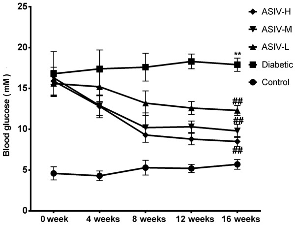
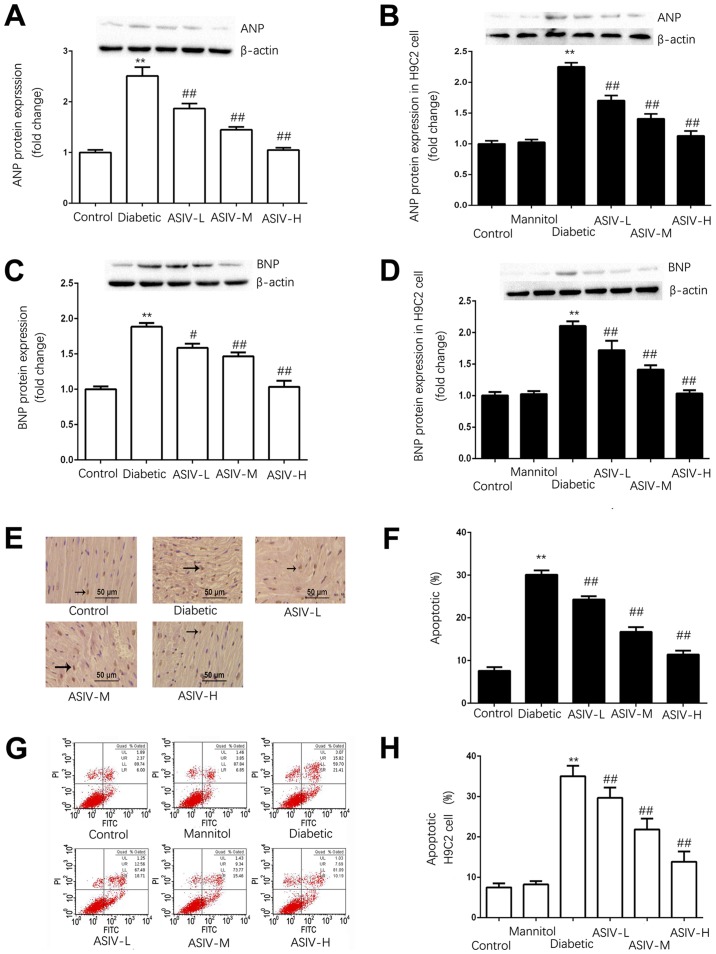
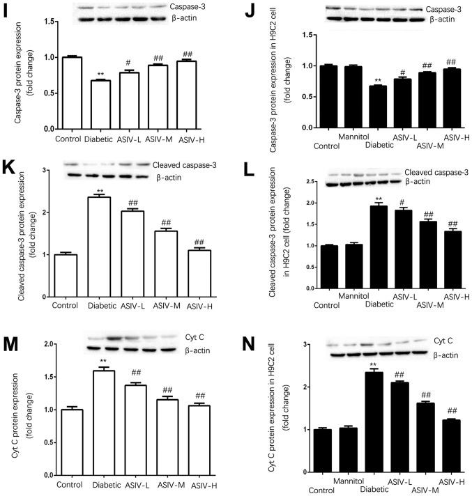
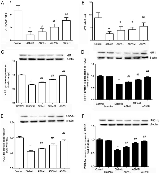
The aim of the present study was to evaluate the protective effect and mechanism of Astragaloside IV (ASIV) on myocardial injury induced by type 2 diabetes, with a focus on energy metabolism. Blood glucose, the hemodynamic index, left ventricular weight/heart weight (LVW/HW), the left ventricular systolic pressure (LVSP), the left ventricular end diastolic pressure (LVEDP) and cell survival rate were measured in streptozotocin‑induced diabetes model rats. Western blot analysis, PCR, hematoxylin‑eosin and TUNEL staining, flow cytometry and ELISA were used to detect: i) Cardiomyocyte damage indicators such as atrial natriuretic peptide (ANP), brain natriuretic peptide (BNP), cytochrome c (Cyt C), caspase‑3, cleaved caspase‑3 and the apoptotic rate; ii) energy metabolism indicators such as ATP/AMP and ADP/AMP; and iii) energy metabolism associated pathway proteins such as peroxisome proliferator‑activated receptor γ coactivator 1‑α (PGC‑1α) and nuclear respiratory factor 1 (NRF1). The present demonstrated increased blood glucose, LVW/HW, LVSP, LVEDP and the cardiomyocyte damage indicators (ANP, BNP, Cyt C and caspase‑3), in the diabetic and high glucose‑treated groups, which were decreased by ASIV. The expression of NRF‑1 and PGC‑1α significantly changed in the model group and was markedly improved following ASIV treatment. Furthermore, the abnormal energy metabolism in the model group was reversed by ASIV. According to the results, ASIV can regulate energy metabolism by regulating the release of PGC‑1α and NRF1 to rescue the abnormal energy metabolism caused by diabetes mellitus, thus decreasing the myocardial damage caused by diabetic cardiomyopathy.


Figures

Figure 1.
Figure 1.
ASIV inhibits the elevation of blood glucose in STZ-induced diabetic rats. Data are expressed as the means ± SD, n=10, **P<0.01 vs. control group. ##P<0.01 vs. diabetic group. ASIV, astragaloside IV.
Figure 2.
Figure 2.
ASIV improves the physiological indexes of STZ-induced diabetic rats and decreases the effects of diabetes on tissue structures. (A) LVEDP and LVSP, (B) ± dp/dtmax and (C) LVW/HW of rats in the different experimental groups. n=10. (D) Hematoxylin and eosin staining in cardiac tissue sections of each group. The black arrow indicates myocardial fibers. (E) Cell survival of H9C2 cells determined by MTT assay. n=8 Data are expressed as the mean ± SD. **P<0.01 vs. the control group. #P<0.05 and ##P<0.01 vs. diabetic group. LVEDP, left ventricular systolic pressure; LVSP, left ventricular end diastolic pressure; ASIV, astragaloside IV; L, low; M, mid; H, high; ± dp/dtmax, maximum left ventricular ascending/descending rate; LVW/HW, ratio of left ventricular weight to heart weight.
Figure 3.
Figure 3.
ASIV decreases the damage of myocardial tissue of diabetic rats and H9C2 cells in high-glucose environment. Protein expression of ANP in (A) the myocardium and (B) H9C2 cells. The protein expression of BNP in (C) the myocardium and (D) H9C2 cells. (E) Myocardial apoptosis assayed by TUNEL staining. TUNEL-positive cells were manifested as a marked appearance of dark brown apoptotic cell nuclei, and (F) the percentage of apoptotic cells in myocardial tissue. (G) The detection of apoptosis in H9C2 cells by AnnexinV-FITC and PI double staining, and (H) the percentage of apoptotic H9C2 cells. The protein expression of (I) caspase-3, (J) cleaved caspase-3 and (K) cytoplasmic Cyt C in the myocardium, and (L) caspase-3, (M) cleaved caspase-3 and (N) cytoplasmic Cyt C in H9C2 cells. The data are expressed as the means ± SD. n=4. **P<0.01 vs. control group. #P<0.05 and ##P<0.01 vs. diabetic group. The black arrows indicate the nucleus of apoptotic cardiomyocytes. ANP, atrial natriuretic peptide; ASIV, astragaloside IV; L, low; M, mid; H, high; BNP, brain natriuretic peptide; PI, propidium iodide; Cyt C, cytochrome c.
Figure 3.
Figure 3.
ASIV decreases the damage of myocardial tissue of diabetic rats and H9C2 cells in high-glucose environment. Protein expression of ANP in (A) the myocardium and (B) H9C2 cells. The protein expression of BNP in (C) the myocardium and (D) H9C2 cells. (E) Myocardial apoptosis assayed by TUNEL staining. TUNEL-positive cells were manifested as a marked appearance of dark brown apoptotic cell nuclei, and (F) the percentage of apoptotic cells in myocardial tissue. (G) The detection of apoptosis in H9C2 cells by AnnexinV-FITC and PI double staining, and (H) the percentage of apoptotic H9C2 cells. The protein expression of (I) caspase-3, (J) cleaved caspase-3 and (K) cytoplasmic Cyt C in the myocardium, and (L) caspase-3, (M) cleaved caspase-3 and (N) cytoplasmic Cyt C in H9C2 cells. The data are expressed as the means ± SD. n=4. **P<0.01 vs. control group. #P<0.05 and ##P<0.01 vs. diabetic group. The black arrows indicate the nucleus of apoptotic cardiomyocytes. ANP, atrial natriuretic peptide; ASIV, astragaloside IV; L, low; M, mid; H, high; BNP, brain natriuretic peptide; PI, propidium iodide; Cyt C, cytochrome c.
Figure 4.
Figure 4.
ASIV regulates the expression of energy metabolism associated signals and the level of energy metabolism in myocardial tissue and H9C2 cells. Ratio of (A) ATP/ADP and (B) ATP/AMP in H9C2 cells. n=8. The protein expression of NRF1 in (C) the myocardium and (D) H9C2 cells. n=4. The protein expression of PGC-1α in (E) the myocardium and (F) H9C2 cells. n=4. The data are expressed as the mean ± SD. **P<0.01 vs. control group. #P<0.05 and ##P<0.01 vs. diabetic group. ASIV, astragaloside IV; L, low; M, mid; H, high; NRF-1, nuclear respiratory factor 1; PGC-1, peroxisome proliferator-activated receptor λ coactivator 1-α.

Similar articles

Cited by

References

    1. Shaw JE, Sicree RA, Zimmet PZ. Global estimates of the prevalence of diabetes for 2010 and 2030. Diabetes Res Clin Pract. 2010;87:4–14. doi: 10.1016/j.diabres.2009.10.007. - DOI - PubMed
    1. Lee CD, Folsom AR, Pankow JS, Brancati FL, Atherosclerosis Risk in Communities (ARIC) Study Investigators Cardiovascular events in diabetic and nondiabetic adults with or without history of myocardial infarction. Circulation. 2004;109:855–860. doi: 10.1161/01.CIR.0000116389.61864.DE. - DOI - PubMed
    1. Knapp M, Tu X, Wu R. Vascular endothelial dysfunction, a major mediator in diabetic cardiomyopathy. Acta Pharmacol Sin. 2019;40:1–8. doi: 10.1038/s41401-018-0042-6. - DOI - PMC - PubMed
    1. Escaned J, Colmenárez H, Ferrer MC, Gutiérrez M, Jiménez-Quevedo P, Hernández R, Alfonso F, Bañuelos C, Deisla LP, Zamorano JL, Macaya C. Diastolic dysfunction in diabetic patients assessed with Doppler echocardiography: Relationship with coronary atherosclerotic burden and microcirculatory impairment. Rev Esp Cardiol. 2009;62:1395–1403. doi: 10.1016/S0300-8932(09)73125-2. - DOI - PubMed
    1. Zheng X, Sun T, Wang X. Activation of type 2 cannabinoid receptors (CB2R) promotes fatty acid oxidation through the SIRT1/PGC-1α pathway. Biochem Biophys Res Commun. 2013;436:377–381. doi: 10.1016/j.bbrc.2013.05.108. - DOI - PubMed

MeSH terms