. 2019 Nov 8;14(11):e0224818.
doi: 10.1371/journal.pone.0224818. eCollection 2019.

Inflammatory cell infiltrates, hypoxia, vascularization, pentraxin 3 and osteoprotegerin in abdominal aortic aneurysms - A quantitative histological study

Affiliations

Inflammatory cell infiltrates, hypoxia, vascularization, pentraxin 3 and osteoprotegerin in abdominal aortic aneurysms - A quantitative histological study

Tereza Blassova et al. PLoS One. .

Abstract

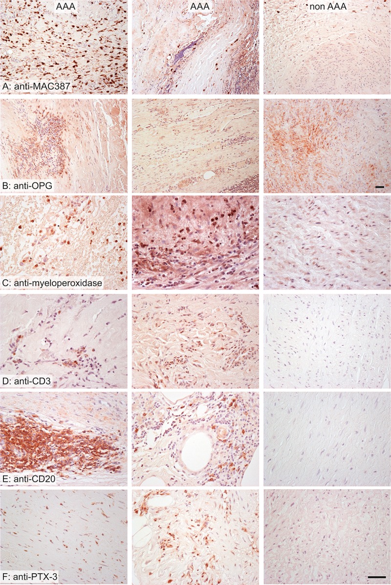
Information about the tissue characteristics of abdominal aortic aneurysms (AAAs), some of which may be reflected in the serum, can help to elucidate AAA pathogenesis and identify new AAA biomarkers. This information would be beneficial not only for diagnostics and follow-up but also for potential therapeutic intervention. Therefore, the aim of our study was to compare the expression of structural proteins, immune factors (T and B lymphocytes, macrophages, neutrophils and pentraxin 3 (PTX3)), osteoprotegerin (OPG), microvessels and hypoxic cells in AAA and nonaneurysmal aortic walls. We examined specimens collected during surgery for AAA repair (n = 39) and from the abdominal aortas of kidney donors without AAA (n = 8). Using histochemical and immunohistochemical methods, we quantified the areas positive for smooth muscle actin, desmin, elastin, collagen, OPG, CD3, CD20, MAC387, myeloperoxidase, PTX3, and hypoxia-inducible factor 1-alpha and the density of CD31-positive microvessels. AAA samples contained significantly less actin, desmin, elastin and OPG, more collagen, macrophages, neutrophils, T lymphocytes, B lymphocytes, hypoxic cells and PTX3, and a greater density of vasa vasorum (VV) than those in non-AAA samples. Hypoxia positively correlated with actin and negatively correlated with collagen. Microvascular density was related to inflammatory cell infiltrates, hypoxia, PTX3 expression and AAA diameter. The lower OPG expression in AAAs supports the notion of its protective role in AAA remodeling. AAA contained altered amounts of structural proteins, implying reduced vascular elasticity. PTX3 was upregulated in AAA and colocalized with inflammatory infiltrates. This evidence supports further evaluation of PTX3 as a candidate marker of AAA. The presence of aortic hypoxia, despite hypervascularization, suggests that hypoxia-induced neoangiogenesis may play a role in AAA pathogenesis. VV angiogenesis of the AAA wall increases its vulnerability.


Conflict of interest statement

The authors have declared that no competing interests exist.

Figures

Fig 1
Fig 1. Wall composition in AAA (left and middle columns) and non-AAA samples (right column).
In the AAA samples, the collagen was more abundant (A), the elastin was partially or mostly destroyed (B), the wall contained more vasa vasorum (D), expressed more hypoxia markers (E) and less contractile phenotype of vascular smooth muscle (C, F). Stained with picrosirius red (A), orcein (B), and immunohistochemistry with anti-smooth muscle actin antibody (C) for visualization of the contractile phenotype of vascular smooth muscle cells, anti-CD31 (D) for visualization of endothelium, anti-HIF 1-alpha (E) for visualization of tissue hypoxia and anti-desmin (F) for visualization of the contractile phenotype of vascular smooth muscle cells; nuclei were stained with Gill’s hematoxylin; scale bar 50 μm.
Fig 2
Fig 2. Wall composition in AAA (left and middle column) and non-AAA samples (right column).
The AAA samples were more infiltrated by macrophages (A), neutrophils (B), T lymphocytes (D) and B lymphocytes (E). The distribution of osteoprotegerin (OPG) was more diffuse in non-AAA (C). The expression of pentraxin 3 was greater in AAA samples (F). Stained immunohistochemistry with anti-MAC387 antibody (A) for visualization of macrophages, anti-osteoprotegerin (B), anti-myeloperoxidase (C) for visualization of neutrophilic granulocytes, anti-CD3 (D) for visualization of T lymphocytes, anti-CD20 (E) for visualization of B lymphocytes, anti-pentraxin 3 (F); nuclei were stained with Gill’s hematoxylin; scale bar 50 μm.
Fig 3
Fig 3. Correlations of the examined histological parameters, in AAA samples.
All correlations significant at p<0.05 are highlighted in boldface. AA(component, space): Area fraction of the respective components within their reference spaces; QA: number of microvessel profiles per section area; int+media: data pooled from the intima and media; wall: data pooled from the wall (i.e., from intima, media and adventitia). Abbreviations of all the examined parameters are explained in S2 Table.
Fig 4
Fig 4. Correlations of the examined histological parameters, in all samples.
All correlations significant at p<0.05 are highlighted in boldface. AA(component, space): Area fraction of the respective components within their reference spaces; QA: number of microvessel profiles per section area; int+media: data pooled from the intima and media; wall: data pooled from the wall (i.e., from intima, media and adventitia). The abbreviations of all the parameters are explained in S2 Table.
Fig 5
Fig 5. Qualitative findings in AAA samples.
A—The basic pattern of the aortic wall was destroyed by newly formed vessels surrounded by inflammatory infiltrates. The elastin fibers (black) were compressed and destroyed. B–The inflammatory cells penetrated among the smooth muscle cells (brown) into the tunica media. C–Lymphocytes (brown) occurred mostly in aggregates resembling lymphoid follicles. D—Rarely, the macrophages (brown) penetrated diffusely the whole wall of the AAA. E–The expression of osteoprotegerin (brown) occurred mostly in the areas infiltrated by leukocytes. F–Similarly, the positivity of pentraxin 3 (brown) also occurred in areas infiltrated by leukocytes. Stained with Verhoeff’s hematoxylin and green trichrome (A) and immunohistochemistry with an antibody against smooth muscle actin (B) for visualization of the contractile phenotype of vascular smooth muscle cells, anti-CD20 (C) for visualization of B lymphocytes, anti-MAC387 (D) for visualization of macrophages, anti-osteoprotegerin (anti-OPG) (E) and anti-pentraxin 3 (anti-PTX3) (F); nuclei were stained with Gill’s hematoxylin; scale bar 100 μm (A, B), 500 μm (C, D) and 50 μm (E, F).

References

    1. Kühnl A, Erk A, Trenner M, Salvermoser M, Schmid V, Eckstein H-H. Incidence, Treatment and Mortality in Patients with Abdominal Aortic Aneurysms: An Analysis of Hospital Discharge Data from 2005–2014. Dtsch Aerzteblatt Online. 2017; 10.3238/arztebl.2017.0391 - DOI - PMC - PubMed
    1. Eberlová L, Tonar Z, Witter K, Křížková V, Nedorost L, Korabečná M, et al. Asymptomatic Abdominal Aortic Aneurysms Show Histological Signs of Progression: a Quantitative Histochemical Analysis. Pathobiol J Immunopathol Mol Cell Biol. 2013;80: 11–23. 10.1159/000339304 - DOI - PubMed
    1. Billaud M, Hill JC, Richards TD, Gleason TG, Phillippi JA. Medial Hypoxia and Adventitial Vasa Vasorum Remodeling in Human Ascending Aortic Aneurysm. Front Cardiovasc Med. 2018;5 10.3389/fcvm.2018.00124 - DOI - PMC - PubMed
    1. Rodella LF, Rezzani R, Bonomini F, Peroni M, Cocchi MA, Hirtler L, et al. Abdominal Aortic Aneurysm and Histological, Clinical, Radiological Correlation. Acta Histochem. 2016;118: 256–262. 10.1016/j.acthis.2016.01.007 - DOI - PubMed
    1. Tanaka H, Zaima N, Sasaki T, Sano M, Yamamoto N, Saito T, et al. Hypoperfusion of the Adventitial Vasa Vasorum Develops an Abdominal Aortic Aneurysm. PloS One. 2015;10: e0134386 10.1371/journal.pone.0134386 - DOI - PMC - PubMed

Publication types