Multicenter Study
. 2019 Nov 7;10(11):899.
doi: 10.3390/genes10110899.

NOP53 as A Candidate Modifier Locus for Familial Non-Medullary Thyroid Cancer

Affiliations
Multicenter Study

NOP53 as A Candidate Modifier Locus for Familial Non-Medullary Thyroid Cancer

Aida Orois et al. Genes (Basel). .

Abstract

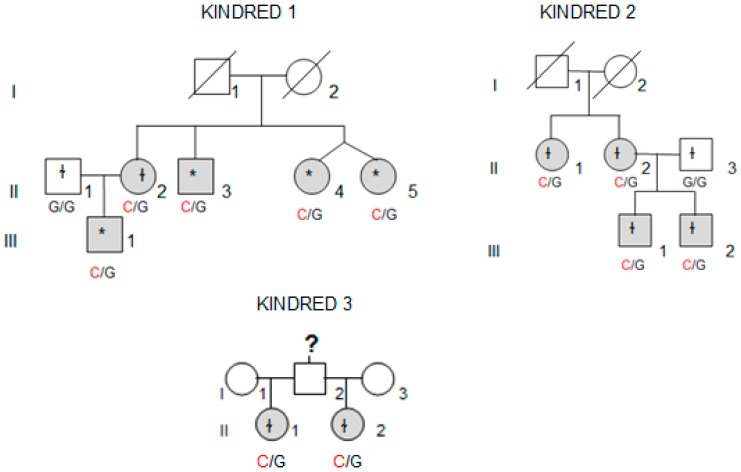
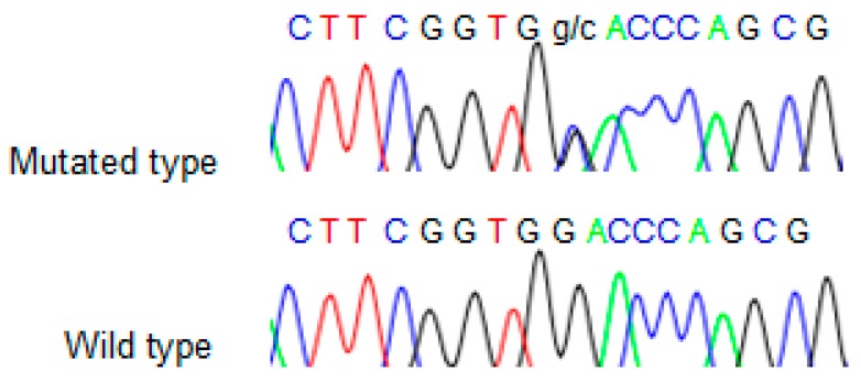
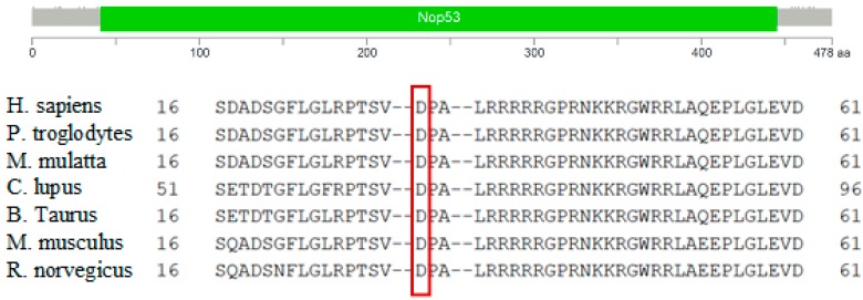
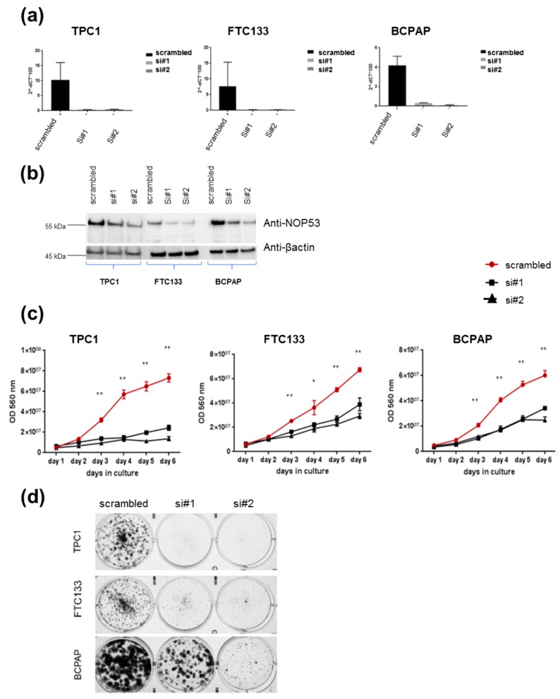
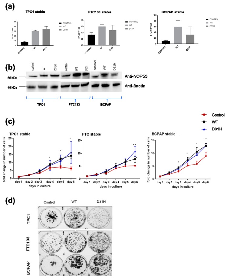
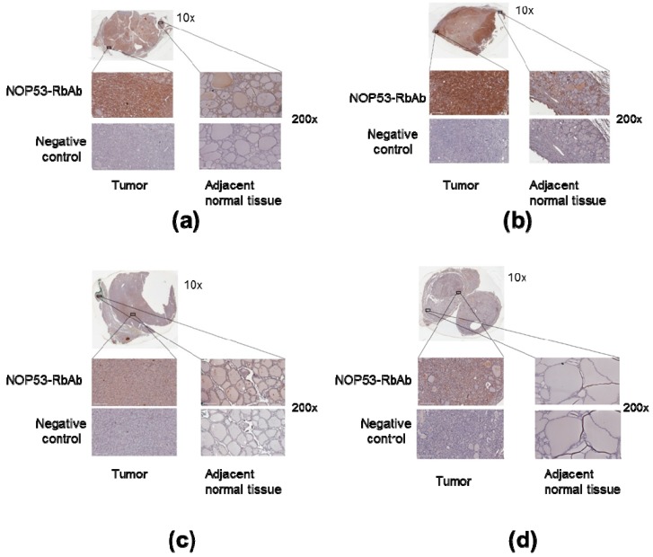
Nonsyndromic familial non-medullary thyroid cancer (FNMTC) represents 3-9% of thyroid cancers, but the susceptibility gene(s) remain unknown. We designed this multicenter study to analyze families with nonsyndromic FNMTC and identify candidate susceptibility genes. We performed exome sequencing of DNA from four affected individuals from one kindred, with five cases of nonsyndromic FNMTC. Single Nucleotide Variants, and insertions and deletions that segregated with all the affected members, were analyzed by Sanger sequencing in 44 additional families with FNMTC (37 with two affected members, and seven with three or more affected members), as well as in an independent control group of 100 subjects. We identified the germline variant p. Asp31His in NOP53 gene (rs78530808, MAF 1.8%) present in all affected members in three families with nonsyndromic FNMTC, and not present in unaffected spouses. Our functional studies of NOP53 in thyroid cancer cell lines showed an oncogenic function. Immunohistochemistry exhibited increased NOP53 protein expression in tumor samples from affected family members, compared with normal adjacent thyroid tissue. Given the relatively high frequency of the variant in the general population, these findings suggest that instead of a causative gene, NOP53 is likely a low-penetrant gene implicated in FNMTC, possibly a modifier.

Keywords: NOP53; genetic abnormalities; molecular testing; oncogenic mutations; thyroid cancer.


Conflict of interest statement

The authors declare no conflict of interest. The funders had no role in the design of the study; in the collection, analyses, or interpretation of data; in the writing of the manuscript, or in the decision to publish the results.

Figures

Figure 1
Figure 1
Family pedigrees for Kindreds 1, 2 and 3 and the NOP53 genotype for each heterozygous mutation (c.91G > C, p. Asp31His). Patients affected by thyroid cancer are shown in grey. The asterisk indicates p. Asp31His variant was observed in whole-exome sequencing (WES) and validated by Sanger sequencing, whereas ɬ indicates that the variant was identified using direct Sanger sequencing.
Figure 2
Figure 2
Sequence obtained from Sanger sequencing from one representative wild type and mutant sample.
Figure 3
Figure 3
Protein domain architecture of NOP53 (GLTSCR2) and conservation of the p.Asp31 position across species. The red frame highlits the amino acid aspartic acid (D) at position 31.
Figure 4
Figure 4
Knockdown of wild-type NOP53 reduces cell proliferation and clonogenicity: (a) Validation of two different siRNAs (si#1 and si#2) targeting NOP53 gene expression in three different thyroid cancer cell lines (TPC1, FTC133 and BCPAP) using qPCR; (b) Validation of two different siRNAs (si#1 and si#2) targeting NOP53 protein expression in three different thyroid cancer cell lines (TPC1, FTC133 and BCPAP) using Western blots. The total protein lysates used were 25 µg for TPC1 cell line; and 30 µg for FTC133 and BCPAP cell lines. GAPDH and β-actin were used as an internal and loading control for qPCR and Western blot, respectively; (c) Transient knockdown of NOP53 in three different cell lines with two siRNAs significantly reduced cell proliferation compared to negative control (scrambled), suggesting a proto-oncogenic function of NOP53; (d) Transient knockdown of NOP53 in three different cell lines with two siRNAs significantly reduced cell clonogenicity compared to negative control (scrambled), suggesting a proto-oncogenic function of NOP53. * indicates adjusted p value < 0.05 compared to control. ** indicate adjusted p value < 0.01 compared to control. Error bars indicate standard deviation.
Figure 5
Figure 5
Effects of stable overexpression of NOP53 in three cell lines (TPC1, FTC133, BCPAP): (a) Validation of stable overexpression of wild type (WT) and D31H mutant NOP53 in three cell lines by qPCR; (b) Validation by Western blot. The lower band corresponds to the endogenous protein expression, whereas the upper band represents the exogenous overexpressed protein. The total protein lysates used were 25 µg for TPC1 cell line, and 30 µg for FTC133 and BCPAP cell lines. GAPDH and β-actin were used as an internal and loading control for qPCR and Western blot, respectively; (c) Overexpression of WT and D31H mutant NOP53 significantly increased the cell proliferation in thyroid cancer cell lines compared to the vector control; (d) Overexpression of WT and D31H mutant NOP53 significantly increased the cell clonogenicity in thyroid cancer cell lines compared to the vector control. * indicates adjusted p value < 0.05 compared to control. ** indicate adjusted p value < 0.01 compared to control. Error bars indicate standard deviation.
Figure 6
Figure 6
Overexpression of NOP53 in tumors from patients with FNMTC. Panels A through D show representative immunohistochemical staining for NOP53 in thyroid cancer samples from the four affected members of Kindred 2: (a) Corresponds to Patient III.1.; (b) Patient II.2.; (c) Patient III.2.; and (d) Patient II.1. Each panel contains an inlet (zoom 10×); two separate regions—from tumor tissue and adjacent normal thyroid tissue—of higher magnification images (zoom 200×); and two higher magnification images (zoom 200×) from a negative control specimen at a similar location. The top left represents tumor staining with NOP53-RbAb, the top right shows adjacent normal tissue staining with NOP53-RbAb, and the two bottom images are negative controls. We observed that the tumor tissue showed a higher expression of NOP53 compared to the adjacent normal thyroid tissue in the four patients studied.

References

    1. Sahasrabudhe R., Stultz J., Williamson J., Lott P., Estrada A., Bohorquez M., Palles C., Polanco-Echeverry G., Jaeger E., Martin L., et al. The HABP2 G534E variant is an unlikely cause of familial non-medullary thyroid cancer. J. Clin. Endocrinol. Metab. 2016;10:1098–1103. doi: 10.1210/jc.2015-3928. - DOI - PMC - PubMed
    1. Moses W., Weng J., Kebebew E. Prevalence, clinicopathologic features, and somatic genetic mutation profile in familial versus sporadic nonmedullary thyroid cancer. Thyroid. 2011;21:367–371. doi: 10.1089/thy.2010.0256. - DOI - PMC - PubMed
    1. Rowland K.J., Moley J.F. Hereditary thyroid cancer syndromes and genetic testing. J. Surg. Oncol. 2015;111:51–60. doi: 10.1002/jso.23769. - DOI - PubMed
    1. Kebebew E. Hereditary non-medullary thyroid cancer. World J. Surg. 2008;32:678–682. doi: 10.1007/s00268-007-9312-z. - DOI - PubMed
    1. Vriens M.R., Suh I., Moses W., Kebebew E. Clinical features and genetic predisposition to hereditary non-medullary thyroid cancer. Thyroid. 2009;19:1343–1349. doi: 10.1089/thy.2009.1607. - DOI - PubMed

Publication types

Substances