. 2019 Nov 8;14(1):40.
doi: 10.1186/s13024-019-0339-z.

Reduced sphingolipid hydrolase activities, substrate accumulation and ganglioside decline in Parkinson's disease

Affiliations

Reduced sphingolipid hydrolase activities, substrate accumulation and ganglioside decline in Parkinson's disease

Mylene Huebecker et al. Mol Neurodegener. .

Erratum in

Abstract

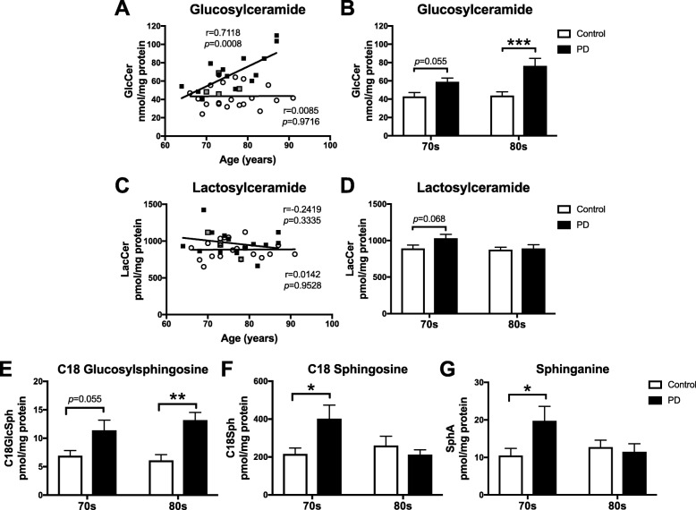
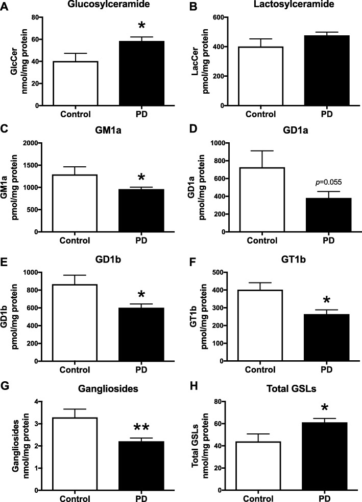
Background: Haploinsufficiency in the Gaucher disease GBA gene, which encodes the lysosomal glucocerebrosidase GBA, and ageing represent major risk factors for developing Parkinson's disease (PD). Recently, more than fifty other lysosomal storage disorder gene variants have been identified in PD, implicating lysosomal dysfunction more broadly as a key risk factor for PD. Despite the evidence of multiple lysosomal genetic risks, it remains unclear how sphingolipid hydrolase activities, other than GBA, are altered with ageing or in PD. Moreover, it is not fully known if levels of glycosphingolipid substrates for these enzymes change in vulnerable brain regions of PD. Finally, little is known about the levels of complex gangliosides in substantia nigra which may play a significant role in ageing and PD.

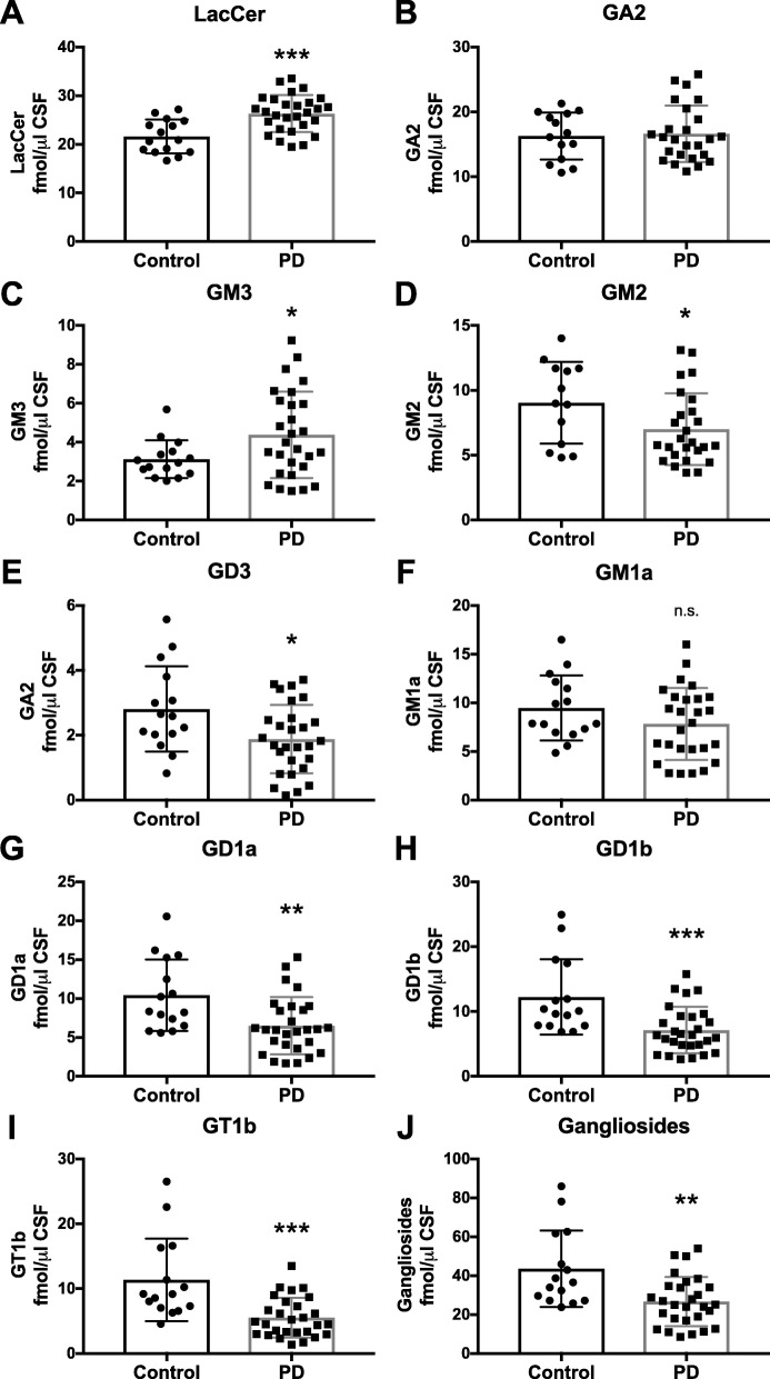
Methods: To study sphingolipid hydrolase activities and glycosphingolipid expression in ageing and in PD, two independent cohorts of human substantia nigra tissues were obtained. Fluorescent 4-methylumbelliferone assays were used to determine multiple enzyme activities. The lysosomal GBA and non-lysosomal GBA2 activities were distinguished using the inhibitor NB-DGJ. Sensitive and quantitative normal-phase HPLC was performed to study glycosphingolipid levels. In addition, glycosphingolipid levels in cerebrospinal fluid and serum were analysed as possible biomarkers for PD.

Results: The present study demonstrates, in two independent cohorts of human post-mortem substantia nigra, that sporadic PD is associated with deficiencies in multiple lysosomal hydrolases (e.g. α-galactosidase and β-hexosaminidase), in addition to reduced GBA and GBA2 activities and concomitant glycosphingolipid substrate accumulation. Furthermore, the data show significant reductions in levels of complex gangliosides (e.g. GM1a) in substantia nigra, CSF and serum in ageing, PD, and REM sleep behaviour disorder, which is a strong predictor of PD.

Conclusions: These findings conclusively demonstrate reductions in GBA activity in the parkinsonian midbrain, and for the first time, reductions in the activity of several other sphingolipid hydrolases. Furthermore, significant reductions were seen in complex gangliosides in PD and ageing. The diminished activities of these lysosomal hydrolases, the glycosphingolipid substrate accumulation, and the reduced levels of complex gangliosides are likely major contributors to the primary development of the pathology seen in PD and related disorders with age.

Keywords: Ageing; Ganglioside; Glucocerebrosidase; Glycosphingolipid; Lysosome; Neurodegeneration; Parkinson’s disease.


Conflict of interest statement

The authors declare that they have no competing interests.

Figures

Fig. 1
Fig. 1
Reduced GBA and GBA2 activities in substantia nigra of PD patients and with normal ageing. GBA and GBA2 β-glucosidase activities were measured using artificial 4-MU-substrate and the inhibitor NB-DGJ. Activity of GBA (a,b) and GBA2 (c,d) were determined in substantia nigra from control subjects and PD patients. PD patients identified as GBA mutation carriers are shown in grey (a,c). Data were analysed using Pearson correlation analysis (a,c) (n = 10 per group) and 2-way ANOVA (b,d) (n = 5 per cohort; * = p < 0.05, ** = p < 0.01, *** = p < 0.001, **** = p < 0.0001). All p-values can be found in the Additional file 3. Bar graphs are presented as mean ± SEM
Fig. 2
Fig. 2
Reduced lysosomal hydrolase activities in substantia nigra of PD patients. Lysosomal hydrolase activities were measured using artificial 4-MU-substrates. Activity of α-galactosidase (a,b), β-hexosaminidase (c,d), β-galactosidase (e,f) and neuraminidase (g,h) were determined in substantia nigra from control subjects and PD patients. PD patients identified as GBA mutation carriers are shown in grey (a,c,e,g). Data were analysed using Pearson correlation analysis (a,c,e,g) (n = 10 per group) and 2-way ANOVA (b,d,f,h) (n = 5 per cohort; * = p < 0.05, ** = p < 0.01, *** = p < 0.001, **** = p < 0.0001). All p-values can be found in the Additional file 3. Bar graphs are presented as mean ± SEM
Fig. 3
Fig. 3
Glucosylceramide and glucosylsphingosine levels are increased in the substantia nigra of PD patients. a-d Substantia nigra from control subjects (n = 20) and PD patients (n = 18) were used to determine glucosylceramide (GlcCer) and lactosylceramide (LacCer) levels with NP-HPLC. a, c Data were analysed using Pearson correlation analysis. b, d Comparison of GlcCer and LacCer levels in 70s-cohorts and 80s-cohorts of control subjects and PD patients was performed using 2-way ANOVA (n = 8–10 per cohort, *** = p < 0.001). PD patients identified as GBA mutation carriers are shown in grey (a,c). e-g Substantia nigra from control subjects and PD patients were used to determine glucosylsphingosine (GlcSph), sphingosine (Sph) and sphinganine (SphA) levels with RP-HPLC. e GlcSph levels in 70s-cohorts and 80s-cohorts of control subjects and PD patients (n = 5 per cohort, ** = p < 0.01, 2-way ANOVA). f Sph levels in 70s-cohorts and 80s-cohorts of control subjects and PD patients (n = 5 per cohort, * = p < 0.05, 2-way ANOVA). g SphA levels in 70s-cohorts and 80s-cohorts of control subjects and PD patients (n = 5 per cohort, * = p < 0.05, 2-way ANOVA). Bar graphs are presented as mean ± SEM
Fig. 4
Fig. 4
Loss of gangliosides in substantia nigra of PD patients and with normal ageing. Levels of GM1a (a,b), GD1a (c,d), GD1b (e,f) and GT1b (g,h) were determined in substantia nigra from control subjects and PD patients with NP-HPLC. Data were analysed using Pearson correlation analysis (a,c,e,g) (n = 18–20 per group) and 2-way ANOVA (b,d,f,h) (n = 8–10 per cohort; * = p < 0.05). i Pearson correlation analysis of the sum of GM1a + GD1a + GD1b + GT1b levels in substantia nigra from control subjects (n = 20) and PD patients (n = 18). j Comparison of ganglioside levels in 70s-cohort vs. 80s-cohort of control subjects and PD patients (n = 8–10 per cohort, ** = p < 0.01, 2-way ANOVA). k Pearson correlation analysis of the sum of GlcCer + LacCer + GM1a + GD1a + GD1b + GT1b levels in substantia nigra from control subjects (n = 20) and PD patients (n = 18) shows that PD is associated with increased GSL load with age. i Comparison of total GSL levels in 70s-cohorts vs. 80s-cohorts of control subjects and PD patients (n = 8–10 per cohort, *** = p < 0.001, 2-way ANOVA). PD patients identified as GBA mutation carriers are shown in grey (a,c,e,g,i,k). Bar graphs are presented as mean ± SEM
Fig. 5
Fig. 5
Reduced lysosomal hydrolase activities in substantia nigra from a second cohort of PD patients. Lysosomal hydrolase activities were measured using artificial 4-MU-substrates. Activity of GBA (a), GBA2 (b), α-galactosidase (c), β-hexosaminidase (d), β-galactosidase (e) and neuraminidase (f) were determined in substantia nigra from age-matched control subjects (n = 5) and PD patients (n = 9). Data were analysed using unpaired t-test (* = p < 0.05, ** = p < 0.01). Data are presented as mean ± SEM
Fig. 6
Fig. 6
Increase in glucosylceramide and loss of gangliosides in substantia nigra from a second cohort of PD patients. Substantia nigra from control subjects (n = 5) and PD patients (n = 20) were used to determine GlcCer (a), LacCer (b) GM1a (c), GD1a (d), GD1b (e) and GT1b (f) levels with NP-HPLC (* = p < 0.05, unpaired t-test). g Comparison of total ganglioside levels (sum of GM1a, GD1a, GD1b and GT1b) in substantia nigra from control subjects and PD patients (** = p < 0.01, unpaired t-test). h Total GSL levels (sum of GlcCer + LacCer + GM1a + GD1a + GD1b + GT1b levels) in substantia nigra from control subjects and PD patients (* = p < 0.05, unpaired t-test). Data are presented as mean ± SEM
Fig. 7
Fig. 7
Significant increase in LacCer and GM3 levels, but significant decrease in complex ganglioside levels in CSF of PD patients. Ante-mortem CSF from control subjects (n = 15) and age-matched PD patients (n = 28) was used to determine LacCer (a), GA2 (b), GM3 (c), GM2 (d), GD3 (e), GM1a (f), GD1a (g), GD1b (h), and GT1b (i) levels with NP-HPLC (* = p < 0.05, ** = p < 0.01, *** = p < 0.001, unpaired t-test). j Total ganglioside levels (sum of GM1a, GD1a, GD1b and GT1b) in ante-mortem CSF from control subjects and PD patients (** = p < 0.01, unpaired t-test). Data are presented as mean ± SD
Fig. 8
Fig. 8
Significant reduction in GM1a and GD1a levels in serum from PD patients and significant reduction in all measured glycosphingolipids, except GlcCer, in serum from RBD patients. Levels of GlcCer (a), LacCer (b), Gb3 (c), Gb4 (d), GM3 (e), GM2 (f), GM1a (g) and GD1a (h) were determined in serum samples from control subjects (n = 15), PD patients (n = 30) and age-matched RBD patients (n = 30) with NP-HPLC (* = p < 0.05, ** = p < 0.01, *** = p < 0.001, **** = p < 0.0001, one-way ANOVA). Data are presented as mean ± SD

References

    1. Kalia LV, Lang AE. Parkinson's disease. Lancet. 2015;386(9996):896–912. doi: 10.1016/S0140-6736(14)61393-3. - DOI - PubMed
    1. Poewe W, Seppi K, Tanner CM, Halliday GM, Brundin P, Volkmann J, et al. Parkinson disease. Nat Rev Dis Primers. 2017;3:17013. doi: 10.1038/nrdp.2017.13. - DOI - PubMed
    1. Engelender S, Isacson O. The threshold theory for Parkinson's disease. Trends Neurosci. 2017;40(1):4–14. doi: 10.1016/j.tins.2016.10.008. - DOI - PubMed
    1. Platt FM. Sphingolipid lysosomal storage disorders. Nature. 2014;510(7503):68–75. doi: 10.1038/nature13476. - DOI - PubMed
    1. Platt FM, d'Azzo A, Davidson BL, Neufeld EF, Tifft CJ. Lysosomal storage diseases. Nat Rev Dis Primers. 2018;4(1):27. doi: 10.1038/s41572-018-0025-4. - DOI - PubMed

Publication types