. 2019 Nov 8;20(1):249.
doi: 10.1186/s12931-019-1223-7.

YY1 mediates TGF-β1-induced EMT and pro-fibrogenesis in alveolar epithelial cells

Affiliations

YY1 mediates TGF-β1-induced EMT and pro-fibrogenesis in alveolar epithelial cells

Chuyi Zhang et al. Respir Res. .

Abstract

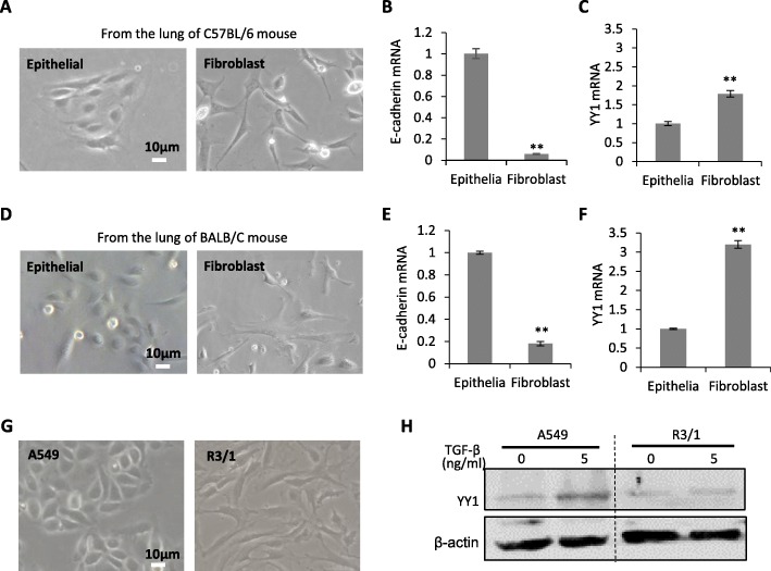
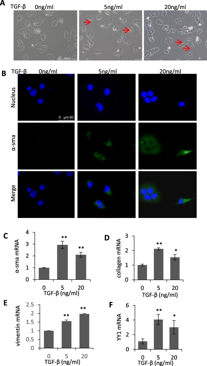
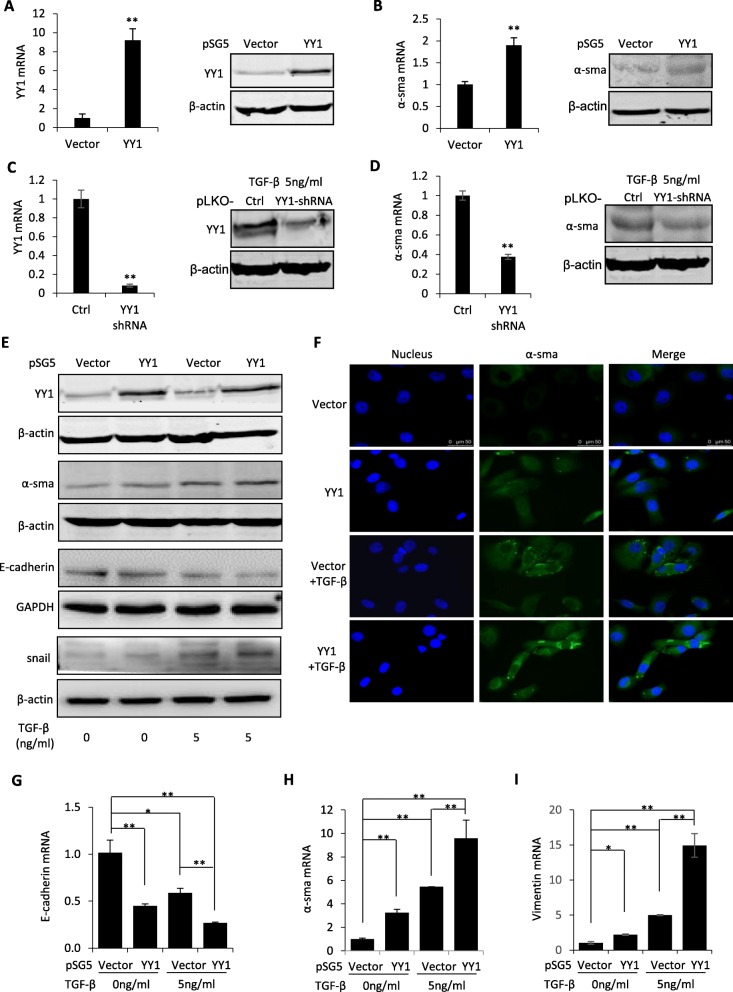
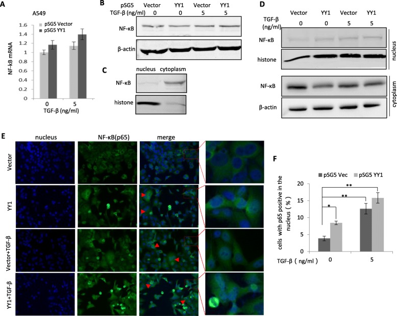
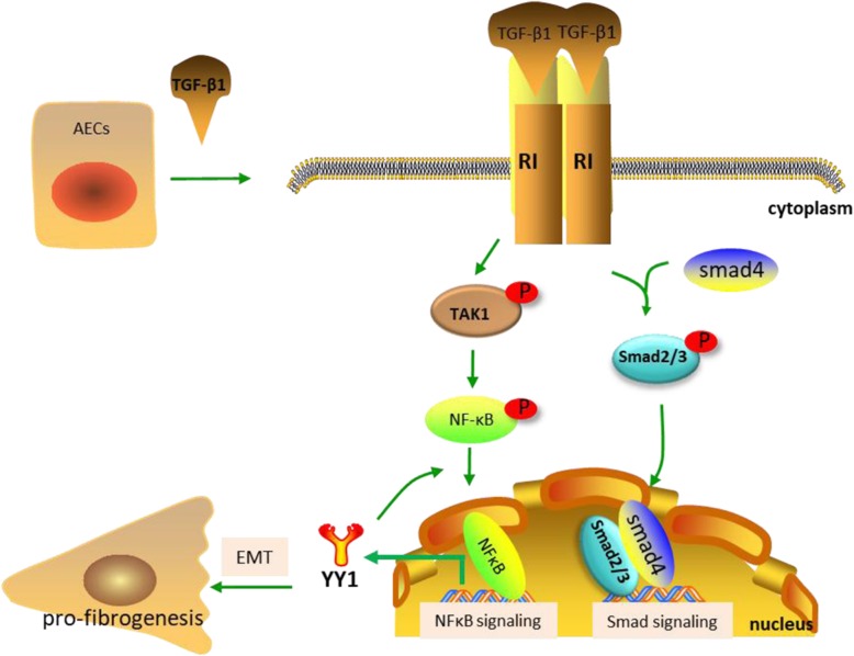
Pulmonary fibrosis is a chronic, progressive lung disease associated with lung damage and scarring. The pathological mechanism causing pulmonary fibrosis remains unknown. Emerging evidence suggests prominent roles of epithelial-mesenchymal transition (EMT) of alveolar epithelial cells (AECs) in myofibroblast formation and progressive pulmonary fibrosis. Our previous work has demonstrated the regulation of YY1 in idiopathic pulmonary fibrosis and pathogenesis of fibroid lung. However, the specific function of YY1 in AECs during the pathogenesis of pulmonary fibrosis is yet to be determined. Herein, we found the higher level of YY1 in primary fibroblasts than that in primary epithelial cells from the lung of mouse. A549 and BEAS-2B cells, serving as models for type II alveolar pulmonary epithelium in vitro, were used to determine the function of YY1 during EMT of AECs. TGF-β-induced activation of the pro-fibrotic program was applied to determine the role YY1 may play in pro-fibrogenesis of type II alveolar epithelial cells. Upregulation of YY1 was associated with EMT and pro-fibrotic phenotype induced by TGF-β treatment. Targeted knockdown of YY1 abrogated the EMT induction by TGF-β treatment. Enforced expression of YY1 can partly mimic the TGF-β-induced pro-fibrotic change in either A549 cell line or primary alveolar epithelial cells, indicating the induction of YY1 expression may mediate the TGF-β-induced EMT and pro-fibrosis. In addition, the translocation of NF-κB p65 from the cytoplasm to the nucleus was demonstrated in A549 cells after TGF-β treatment and/or YY1 overexpression, suggesting that NF-κB-YY1 signaling pathway regulates pulmonary fibrotic progression in lung epithelial cells. These findings will shed light on the better understanding of mechanisms regulating pro-fibrogenesis in AECs and pathogenesis of lung fibrosis.

Keywords: EMT, alveolar epithelial cells; Pulmonary fibrosis; YY1.


Conflict of interest statement

There are no conflicts of interest associated with this study.

Figures

Fig. 1
Fig. 1
YY1 expression in alveolar epithelial cells. a The fibroblast cells and alveolar epithelial cells isolated from the fresh lung of C57BL/6 mouse. Scale bar: 10 μm. b and c Quantitative RT-PCR analysis of E-cadherin (b) and YY1 (c) expression in the fibroblast and epithelial cells in a. d The fibroblast cells and alveolar epithelial cells isolated from the fresh lung of BALB/C mouse. Scale bar: 10 μm. e and f Quantitative RT-PCR analysis of E-cadherin (e) and YY1 (f) expression in the fibroblast and epithelial cells in d. g Morphology of A549 and R3/1 lung epithelial cells. Scale bar: 10 μm. h Western blot analysis of YY1 in A549 and R3/1 cells with or without TGF-β treatment for 24 h. GAPDH served as housekeeping gene for normalization in Quantitative RT-PCR assays. β-actin served as loading control for western blots. Data are presented as mean ± SEM (n = 3), **p < 0.01
Fig. 2
Fig. 2
Upregulation of YY1 was associated with the TGF-β induced EMT and pro-fibrotic changes in A549 cells. a Morphological change of A549 cells induced by different concentrations of TGF-β. The arrows indicated representative profibrotic cells. Scale bar: 200 μm. b Immunofluorescence staining demonstrated the induction of α-sma by 0, 5 and 20 ng/ml of TGF-β treatment. Scale bar: 50 μm. c-f Quantitative RT-PCR analysis of mesenchymal and pro-fibrotic markers including α-sma (c), collagen (d), vimentin (e) and YY1 (f) in A549 cells before or after treatment with 5 and 20 ng/ml of TGF-β. TGF-β was applied for 48 h. Data are presented as mean ± SEM (n = 3). All comparisons were made with TGF-β-treated cells vs untreated cells. *p < 0.05, **p < 0.01
Fig. 3
Fig. 3
YY1 mediated TGF-β-induced EMT and pro-fibrotic phenotypes. a, b QRT-PCR and western blot were applied to analyze YY1 (a) and α-sma (b) expression at the mRNA and protein levels in A549 cells transfected with YY1-overexpressing plasmid or pSG5 vector control. c, d QRT-PCR and western blot were applied to analyze YY1 (c) and α-sma (d) expression at the mRNA and protein levels in A549 cells transfected with YY1-shRNA or pLKO vector control. e Proteomic analyses of α-sma, YY1, E-cadherin and snail in A549 cells treated by 0 or 5 ng/ml of TGF-β with or without overexpression of YY1. f Immunofluorescence staining of α-sma in A549 cells treated by 0 or 5 ng/ml of TGF-β with or without overexpression of YY1. Scale bar: 50 μm. g-i Primary alveolar epithelial cells isolated from the lung of C57BL/6 J mice were applied to perform QRT-PCR analyses on the expression of E-cadherin (g), α-sma (h) and vimentin (i) under treatment of 0 or 5 ng/ml of TGF-β with or without overexpression of YY1. β-actin or GAPDH served as loading control of western blots as indicated. Data are presented as mean ± SEM (n = 3), *p < 0.05, **p < 0.01
Fig. 4
Fig. 4
TGF-β induced translocation of the NF-κB protein from cytoplasm to nucleus. a and b Analyses of NF-κB (p65) at the mRNA level (a) or protein level (b) in A549 cells treated by 0 or 5 ng/ml of TGF-β with or without overexpression of YY1. Quantitative RT-PCR and western blot were used, respectively. c Localization of NF-κB protein in the cytoplasm and the nucleus of A549 cells under normal condition. d Analysis of NF-κB in the cytoplasm and the nucleus of A549 cells treated by 0 or 5 ng/ml of TGF-β with or without overexpression of YY1. Western blot was performed. Histone served as a marker for nucleus lysates. β-actin served as a marker for cytoplasm lysates. e immunofluorescence staining of A549 cells with antibody against the NF-κB p65 protein to indicate distribution of NF-κB in the cytoplasm and the nucleus. Representative cells with relocalization of NF-κB to the nucleus were indicated with arrows in red. f Quantitative analysis of the cells with NF-κB positive in the nucleus in (e). Data are presented as mean ± SEM (n = 3). *p < 0.05, **p < 0.01
Fig. 5
Fig. 5
Schematic representation of the hypothetical mechanism by which YY1 is involved in the TGF-β-induced EMT and pro-fibrotic phenotypes in alveolar epithelial cells

References

    1. Zoz DF, Lawson WE, Blackwell TS. Idiopathic pulmonary fibrosis: a disorder of epithelial cell dysfunction. Am J Med Sci. 2011;341:435–438. - PMC - PubMed
    1. Selman M, Pardo A. Role of epithelial cells in idiopathic pulmonary fibrosis: from innocent targets to serial killers. Proc Am Thorac Soc. 2006;3:364–372. - PubMed
    1. Selman M, Pardo A. Idiopathic pulmonary fibrosis: an epithelial/fibroblastic cross-talk disorder. Respir Res. 2002;3:3. - PMC - PubMed
    1. Hinz B, Phan SH, Thannickal VJ, Galli A, Bochaton-Piallat ML, Gabbiani G. The myofibroblast: one function, multiple origins. Am J Pathol. 2007;170:1807–1816. - PMC - PubMed
    1. Epperly MW, Guo H, Gretton JE, Greenberger JS. Bone marrow origin of myofibroblasts in irradiation pulmonary fibrosis. Am J Respir Cell Mol Biol. 2003;29:213–224. - PubMed

MeSH terms