

The skeletal phenotype of intermediate GM1 gangliosidosis: Clinical, radiographic and densitometric features, and implications for clinical monitoring and intervention
- PMID: 31704340
- PMCID: PMC6937522
- DOI: 10.1016/j.bone.2019.115142
The skeletal phenotype of intermediate GM1 gangliosidosis: Clinical, radiographic and densitometric features, and implications for clinical monitoring and intervention
Abstract
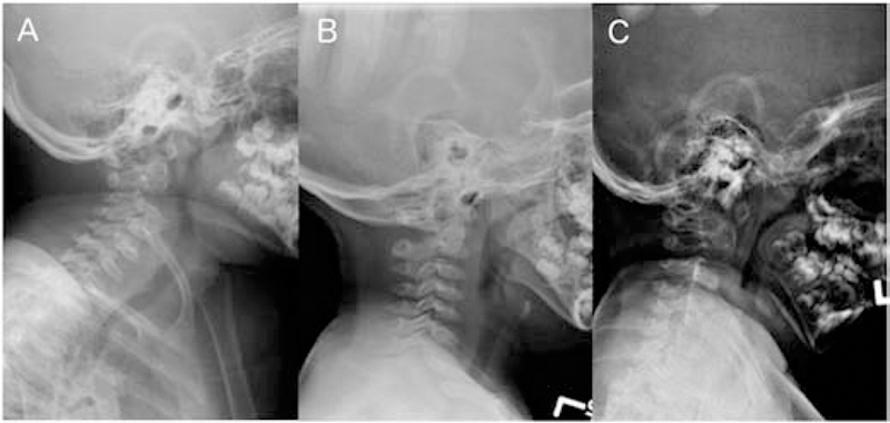
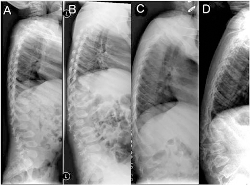
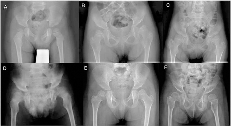
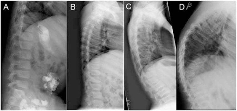
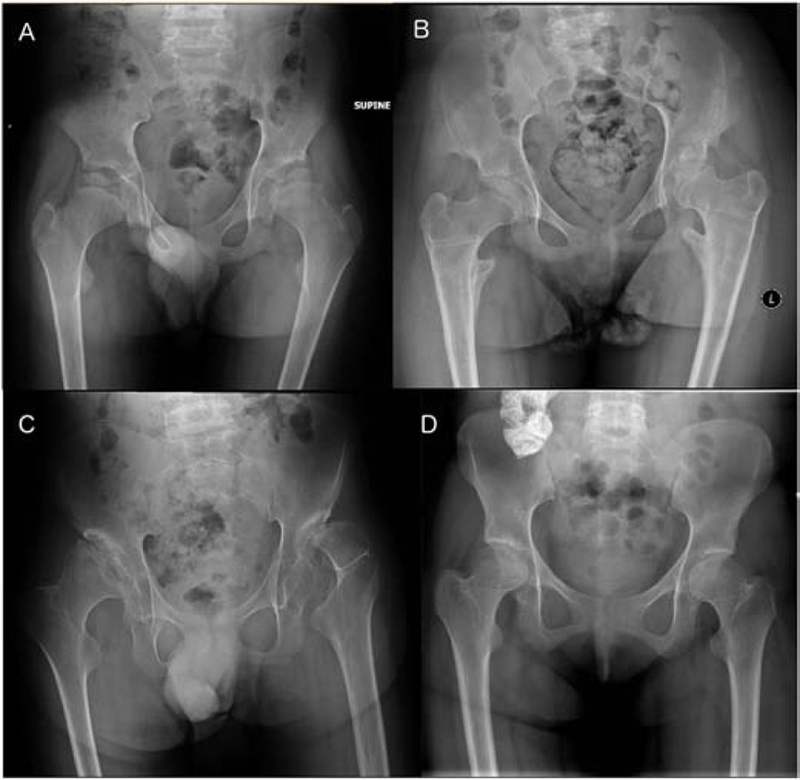
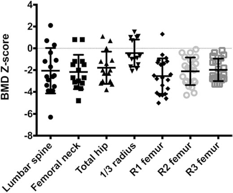
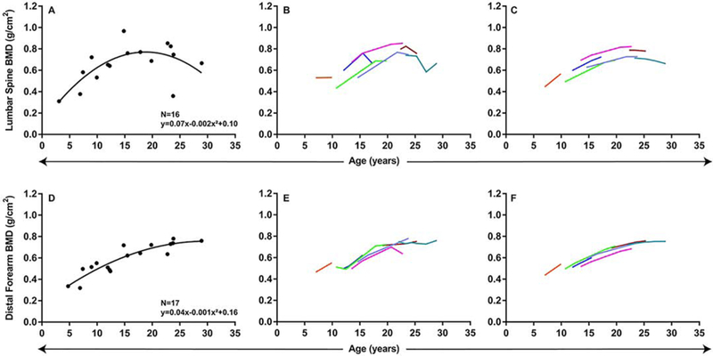
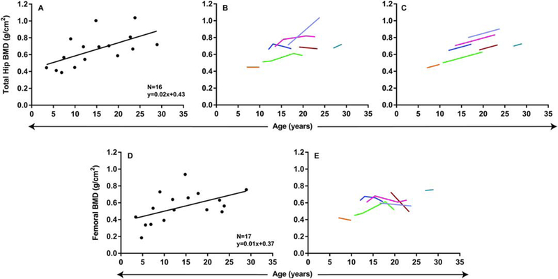
GM1 gangliosidosis is a lysosomal storage disorder caused by mutations in GLB1 encoding a lysosomal β-galactosidase. This disease is a continuum from the severe infantile form with rapid neurological decline to the chronic adult form, which is not life-limiting. The intermediate or type 2 form can be further classified into late infantile and juvenile forms. The frequency and severity of skeletal outcomes in late infantile and juvenile patients have not been characterized. Our goals are to describe the radiological skeletal abnormalities, bone mineral density (BMD), and frequency of fractures in patients with intermediate GM1 gangliosidosis. We evaluated 13 late infantile and 21 juvenile patients as part of an ongoing natural history study. Average time from onset of symptoms to diagnosis was 1.9 and 6.3 years for late infantile and juvenile patients, respectively. All late infantile patients had odontoid hypoplasia and pear-shaped vertebral bodies, the frequency of which was significantly different than in patients with juvenile disease (none and 14%, respectively). Juvenile patients had irregular endplates of the vertebral bodies (15/21), central indentation of endplates (10/21), and squared and flat vertebral bodies (10/21); all allowed radiographic differentiation from late infantile patients. Lumbar spine, femoral neck, and total hip BMD were significantly decreased (-2.1, -2.2, and -1.8 Z-scores respectively). Lumbar spine BMD peaked at 19 years, while distal forearm BMD peaked at 30 years. Despite low BMD, no patients exhibited fractures. We have demonstrated that all late infantile patients have some degree of odontoid hypoplasia suggesting the need for cervical spine evaluation particularly prior to anesthesia, whereas juvenile patients had variable skeletal involvement often affecting activities of daily living. Type 2 GM1 gangliosidosis patients have skeletal abnormalities that are both an early indication of their diagnosis, and require monitoring and management to ensure the highest possible quality of life.
Keywords: Beta-galactosidase deficiency; GM1 gangliosidosis type 2; Lysosomal storage disease; Skeletal dysplasia.
Published by Elsevier Inc.
Figures
Similar articles
-
GM1 Gangliosidosis-A Mini-Review.Front Genet. 2021 Sep 3;12:734878. doi: 10.3389/fgene.2021.734878. eCollection 2021. Front Genet. 2021. PMID: 34539759 Free PMC article. Review.
-
Late-infantile GM1 gangliosidosis: A case report.Medicine (Baltimore). 2022 Jan 7;101(1):e28435. doi: 10.1097/MD.0000000000028435. Medicine (Baltimore). 2022. PMID: 35029890 Free PMC article.
-
Diagnostic challenge for the rare lysosomal storage disease: Late infantile GM1 gangliosidosis.Brain Dev. 2018 May;40(5):383-390. doi: 10.1016/j.braindev.2018.01.009. Epub 2018 Feb 10. Brain Dev. 2018. PMID: 29439846
-
MRI/MRS as a surrogate marker for clinical progression in GM1 gangliosidosis.Am J Med Genet A. 2016 Mar;170(3):634-44. doi: 10.1002/ajmg.a.37468. Epub 2015 Dec 8. Am J Med Genet A. 2016. PMID: 26646981
-
[beta-galactosidosis--GM1 gangliosidosis and Morquio B disease].Nihon Rinsho. 1995 Dec;53(12):2960-6. Nihon Rinsho. 1995. PMID: 8577043 Review. Japanese.
Cited by
-
GM1 Gangliosidosis-A Mini-Review.Front Genet. 2021 Sep 3;12:734878. doi: 10.3389/fgene.2021.734878. eCollection 2021. Front Genet. 2021. PMID: 34539759 Free PMC article. Review.
-
GM1 Gangliosidosis Type II: Results of a 10-Year Prospective Study.medRxiv [Preprint]. 2024 Jan 4:2024.01.04.24300778. doi: 10.1101/2024.01.04.24300778. medRxiv. 2024. Update in: Genet Med. 2024 Jul;26(7):101144. doi: 10.1016/j.gim.2024.101144. PMID: 38313286 Free PMC article. Updated. Preprint.
-
A GM1 gangliosidosis mutant mouse model exhibits activated microglia and disturbed autophagy.Exp Biol Med (Maywood). 2021 Jun;246(11):1330-1341. doi: 10.1177/1535370221993052. Epub 2021 Feb 14. Exp Biol Med (Maywood). 2021. PMID: 33583210 Free PMC article.
-
GM1 gangliosidosis type II: Results of a 10-year prospective study.Genet Med. 2024 Jul;26(7):101144. doi: 10.1016/j.gim.2024.101144. Epub 2024 Apr 16. Genet Med. 2024. PMID: 38641994 Free PMC article.
-
Late-infantile GM1 gangliosidosis: A case report.Medicine (Baltimore). 2022 Jan 7;101(1):e28435. doi: 10.1097/MD.0000000000028435. Medicine (Baltimore). 2022. PMID: 35029890 Free PMC article.
References
-
- Norman RM, Urich H, Tingey AH, Goodbody RA, Tay-Sachs’ disease with visceral involvement and its relationship to Niemann-Pick’s disease, J Pathol Bacteriol 78 (1959) 409–421. - PubMed
-
- Regier DS, Tifft CJ, GLB1-Related Disorders, in: Adam MP, Ardinger HH, Pagon RA, Wallace SE, Bean LJ, Stephens K, Amemiya A (Eds.), GeneReviews®, University of Washington, Seattle, Seattle (WA), 1993. http://www.ncbi.nlm.nih.gov/books/NBK164500/ (accessed January 31, 2019). - PubMed
-
- Suzuki Y, Nanba E, Matsuda J, Higaki K, Oshima A, β-Galactosidase Deficiency (β-Galactosidosis): GM1 Gangliosidosis and Morquio B Disease, in: Beaudet AL, Vogelstein B, Kinzler KW, Antonarakis SE, Ballabio A, Gibson KM, Mitchell G (Eds.), The Online Metabolic and Molecular Bases of Inherited Disease, The McGraw-Hill Companies, Inc., New York, NY, 2014. ommbid.mhmedical.com/content.aspx?aid=1121415439 (accessed February 1, 2019).
-
- Regier DS, Oetgen M, Tanpaiboon P, Mucopolysaccharidosis Type IVA, in: Adam MP, Ardinger HH, Pagon RA, Wallace SE, Bean LJ, Stephens K, Amemiya A (Eds.), GeneReviews®, University of Washington, Seattle, Seattle (WA), 1993. http://www.ncbi.nlm.nih.gov/books/NBK148668/ (accessed February 1, 2019).
Publication types
MeSH terms
Grants and funding
LinkOut - more resources
Full Text Sources
