Review
. 2020 Apr;104(4):451-460.
doi: 10.1136/bjophthalmol-2019-315086. Epub 2019 Nov 8.

Macular dystrophies: clinical and imaging features, molecular genetics and therapeutic options

Affiliations
Review

Macular dystrophies: clinical and imaging features, molecular genetics and therapeutic options

Najiha Rahman et al. Br J Ophthalmol. 2020 Apr.

Abstract

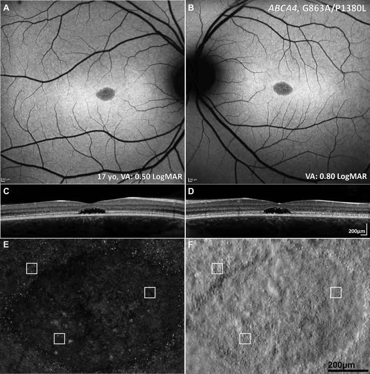
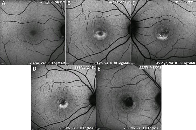
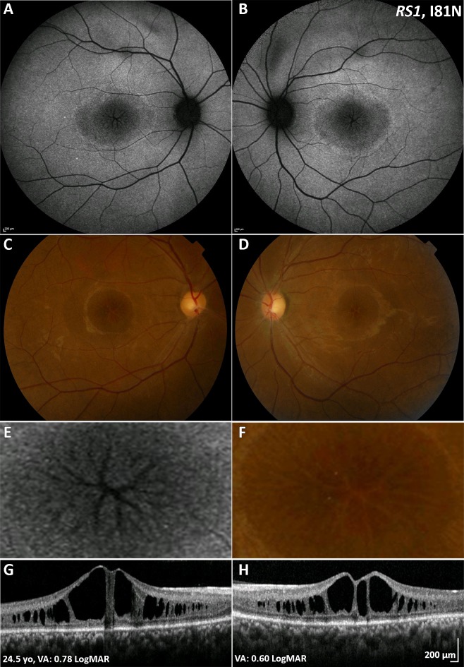
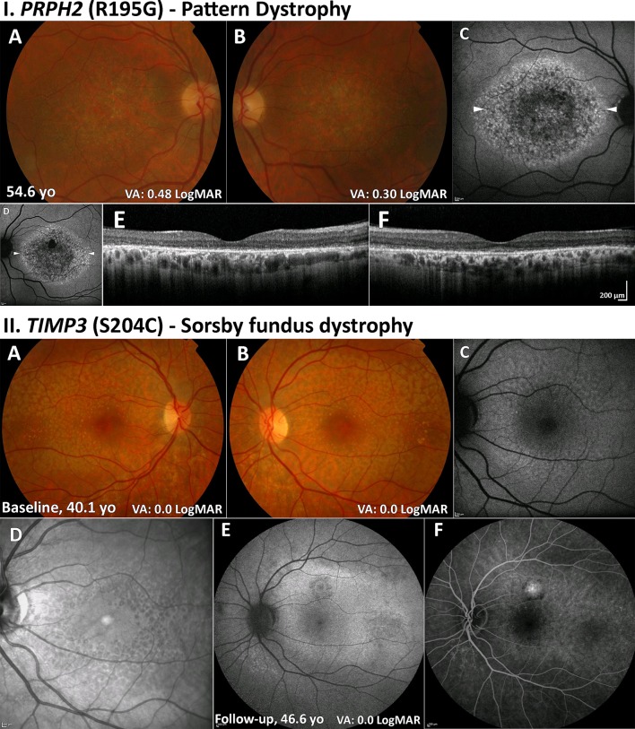
Macular dystrophies (MDs) consist of a heterogeneous group of disorders that are characterised by bilateral symmetrical central visual loss. Advances in genetic testing over the last decade have led to improved knowledge of the underlying molecular basis. The developments in high-resolution multimodal retinal imaging have also transformed our ability to make accurate and more timely diagnoses and more sensitive quantitative assessment of disease progression, and allowed the design of optimised clinical trial endpoints for novel therapeutic interventions. The aim of this review was to provide an update on MDs, including Stargardt disease, Best disease, X-linked r etinoschisis, pattern dystrophy, Sorsby fundus dystrophy and autosomal dominant drusen. It highlights the range of innovations in retinal imaging, genotype-phenotype and structure-function associations, animal models of disease and the multiple treatment strategies that are currently in clinical trial or planned in the near future, which are anticipated to lead to significant changes in the management of patients with MDs.

Keywords: ABCA4; ADD; BEST1; Best disease; EFEMP1; PRPH2; RS1; STGD; Sorsby fundus dystrophy; Stargardt disease; TIMP3; X-linked retinoschisis; XLRS; autosomal dominant drusen; gene therapy; macular dystrophy; pattern dystrophy; pharmacological therapy; retina; stem cells.


Conflict of interest statement

Competing interests: None declared.

Figures

Figure 1
Figure 1
Multimodal imaging of a 16-year-old adolescent with molecularly confirmed STGD. (A, B) Fundus autofluorescence images showing a central area of decreased signal at the macula. (C, D) Corresponding horizontal transfoveal optical coherence tomography scans showing central loss of the ellipsoid zone. (A–D) Findings are symmetrical between the eyes. (E, F) Adaptive optics scanning light ophthalmoscopy of the right eye. Confocal detection (A) and split detection (B) over the foveal lesion in exact coregistration. The white box of 55×55 µm denotes regions of interest in the exact same locations in the two images. Cones are more reliably identified using split detection (B) due to the poor waveguiding of the outer segments in confocal imaging (A). VA, STGD, Stargardt disease; VA, visual acuity.
Figure 2
Figure 2
BD (BEST1) fundus autofluorescence imaging of four patients at different stages. (A) Stage 1: normal previtelliform presentation. (B) Stage 2: vitelliform lesion, classical appearance of a single, symmetrical egg yolk-like lesion at the fovea. (C) Stage 3: pseudohypopyon, material gravitates inferiorly within the vitelliform lesion. (D) Stage 4: vitelliruptive stage, the material ‘scrambles’. (B and D) Images are from the same patient over 4.4 years of follow-up. (E) Stage 5: macular atrophy. BD, Best disease; VA, visual acuity.
Figure 3
Figure 3
XLRS (RS1) multimodal imaging of a 25-year-old patient with XLRS. (A, B) Fundus autofluorescence imaging, with a central decreased signal over the macula and a rim of increased signal, absent signal in a ‘spoke wheel’ pattern over the central fovea in both eyes, shown in greater magnification in (E) for the right eye. (C, D) Colour fundus photographs with the same pattern as (A, B). (F) Greater magnification of the foveal centre of the right eye. (G, H) Transfoveal optical coherence tomography scans showing the extent of schisis cavities in the outer retina. VA, visual acuity; XLRS, X-linked retinoschisis.
Figure 4
Figure 4
PD (PRPH2) and SFD (TIMP3). (I) PD (A, B): colour fundus photographs with bull’s-eye maculopathy-like retinal pigment epithelial changes and fine mottled symmetrical depigmentation of the macula. (C, D) FAF imaging displays a florid speckled appearance with areas of increased and decreased macular autofluorescence. The white arrowheads denote the location of the optical coherence tomography line scans shown in (E) and (F). (E, F) Extensive disruption of the ellipsoid zone and retinal pigment epithelium hypertrophy in both eyes. II. SFD (A, B): colour fundus photographs with fine symmetrical drusen-like deposits at the posterior pole. (C) FAF imaging of the left eye displays patchy ill-defined increased autofluorescence. (D) Infrared image over the same location as (C) readily depicting the drusen-like deposits. (E) FAF image after 6.5 years of follow-up showing a superior area of CNV. (F) Fundus fluorescein angiography showing an inactive CNV. CNV, choroidal neovascularisation; FAF, fundus autofluorescence; PD, pattern dystrophy; SFD, Sorsby fundus dystrophy; VA, visual acuity.
Figure 5
Figure 5
ADD (EFEMP1) multimodal imaging of a 44-year-old patient. (A, B) Colour fundus photographs with characteristic radial distribution of macular drusen. Note the CNV in the right eye (A). (C, D) Fundus autofluorescence images with the drusen associated with an increased signal. The black dashes denote the location of the OCT line scans shown in (E) and (F). (E, F) In both eyes hyper-reflective thickening of the retinal pigment epithelium-Bruch membrane complex, with disrupted photoreceptor integrity. (E) CNV is seen associated with a reduction in VA. ADD, autosomal dominant drusen; CNV, choroidal neovascularisation; EFEMP1, EGF-containing fibulin-like extracellular matrix protein-1; VA, visual acuity.

References

    1. Michaelides M, Hunt DM, Moore AT. The genetics of inherited macular dystrophies. J Med Genet 2003;40:641–50. 10.1136/jmg.40.9.641 - DOI - PMC - PubMed
    1. Scholl HPN, Strauss RW, Singh MS, et al. . Emerging therapies for inherited retinal degeneration. Sci Transl Med 2016;8:368rv6 10.1126/scitranslmed.aaf2838 - DOI - PubMed
    1. Michaelides M, Jeffery G, Moore AT. Developmental macular disorders: phenotypes and underlying molecular genetic basis. Br J Ophthalmol 2012;96:917–24. 10.1136/bjophthalmol-2011-300994 - DOI - PubMed
    1. Schachat AP, Wilkinson CP, Hinton DR, et al. . Ryan's retina. 6th edn Elsevier, 2018.
    1. Khan KN, Kasilian M, Mahroo OAR, et al. . Early patterns of macular degeneration in ABCA4 -associated retinopathy. Ophthalmology 2018;125:735–46. 10.1016/j.ophtha.2017.11.020 - DOI - PMC - PubMed

Publication types