. 2019 Nov 8;9(1):16327.
doi: 10.1038/s41598-019-52808-z.

Evidence of Cardiovascular Calcification and Fibrosis in Pseudoxanthoma Elasticum Mouse Models Subjected to DOCA-Salt Hypertension

Affiliations

Evidence of Cardiovascular Calcification and Fibrosis in Pseudoxanthoma Elasticum Mouse Models Subjected to DOCA-Salt Hypertension

Loukman Omarjee et al. Sci Rep. .

Abstract

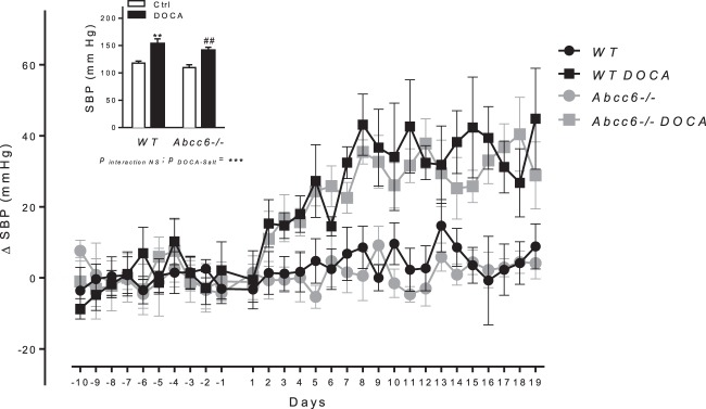
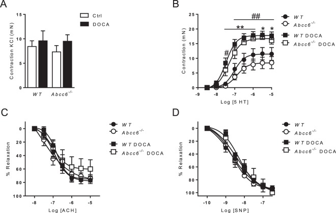
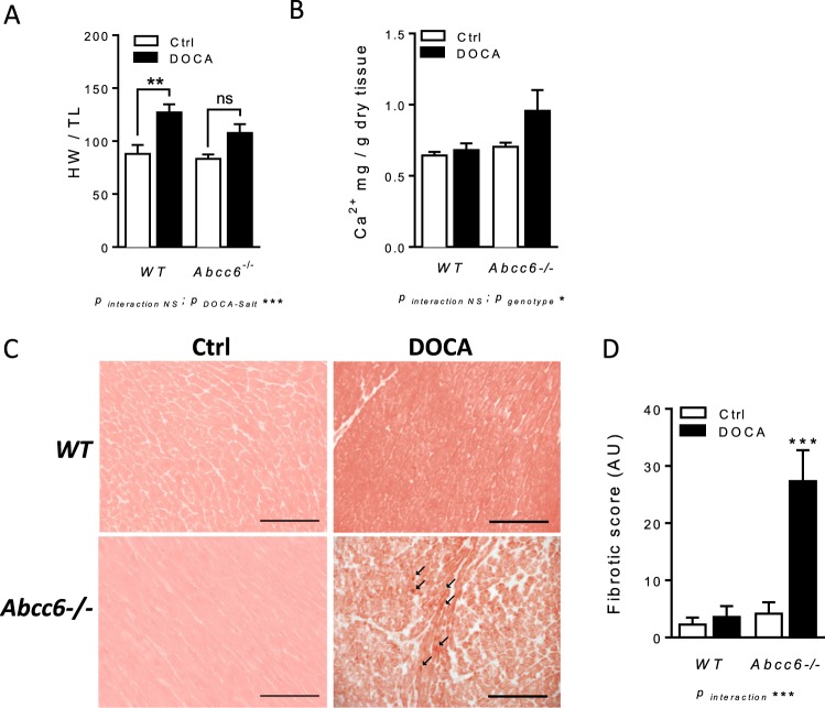
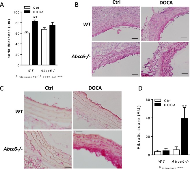
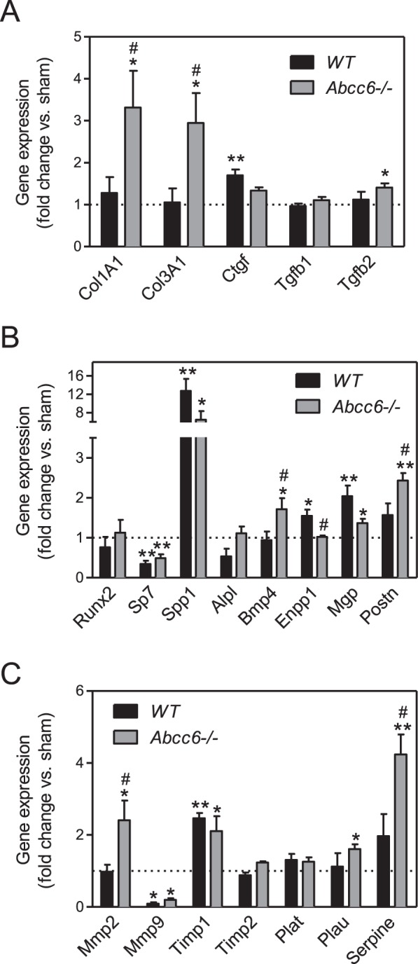
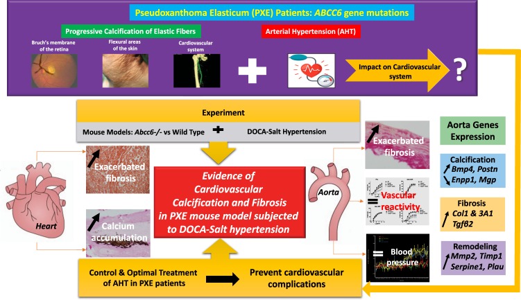
Pseudoxanthoma Elasticum (PXE) is a rare disorder characterized by fragmentation and progressive calcification of elastic fibres in connective tissues. Although arterial hypertension (AHT) has been reported in PXE patients, its impact on pathological manifestations has as yet been unexplored. We investigated the consequences of experimental AHT on Abcc6-/- PXE mouse models. Experimental AHT was induced by deoxycorticosterone acetate (DOCA-salt) in uni-nephrectomised mice. Blood pressure (BP) and vascular reactivity were monitored using tail-cuff plethysmography and myography respectively. Calcium content and fibrosis were assessed using colorimetry, Von Kossa and Sirius red staining respectively. The gene expression implicated in vascular biology was measured using quantitative polymerase chain reaction. DOCA-salt induced a matching rise in BP in Abcc6-/- and WT mice. Aortic ring contraction and relaxation in vitro were comparable. Calcium accumulated in the hearts of hypertensive Abcc6-/- mice along with significant fibrosis in the myocardium and aorta by contrast with the WT mice. In hypertensive Abcc6-/- mouse aortas, these results were corroborated by gene expression patterns favouring calcification, fibrosis and extracellular matrix remodelling. Abcc6 loss-of-function is associated with greater cardiovascular calcification and fibrosis in mice subjected to DOCA-Salt hypertension. These results suggest likely cardiovascular deterioration in PXE patients with AHT, necessitating diligent BP monitoring.


Conflict of interest statement

The authors declare no competing interests.

Figures

Figure 1
Figure 1
Comparable DOCA-salt-induced hemodynamic parameters changes in Abcc6−/− and WT mice. Systolic blood pressure kinetic characteristics as well as mean values over ultimate five days of DOCA-salt treatment. Data represent mean ± SEM of six mice from each group *p < 0.05, **p < 0.01 WT untreated versus DOCA; #p < 0.05, ##p < 0.01 Abcc6−/− untreated versus DOCA.
Figure 2
Figure 2
Comparable DOCA-salt-induced vascular reactivity changes in Abcc6−/− and WT mice. (A) Contraction of aortic rings in response to KCl, (B) Dose-response contraction curve in response to 5-HT and (C,D) endothelium-dependent and -independent relaxation (ACh and SNP, respectively) evaluated using wire myography. Data represent mean ± SEM of six mice from each group *p < 0.05, **p < 0.01 WT untreated versus DOCA; #p < 0.05, ##p < 0.01 Abcc6−/− untreated versus DOCA.
Figure 3
Figure 3
DOCA-salt-induced cardiac remodelling in Abcc6−/− mice: hypertrophy, calcium accumulation and fibrosis. (A) Heart weight normalized to tibia length (HW/TL) (B) Colorimetric determination of total tissue calcium level in left ventricles and (C) representative images and (D) quantification of Sirius red staining. Data represent mean ± SEM of six mice from each group *p < 0.05, **p < 0.01, ***p < 0.001.
Figure 4
Figure 4
DOCA-salt-induced aortic fibrosis excepting hypertrophy in Abcc6−/− mice. (A) Wall thickness measured in TA sections. (B) Representative images of Von Kossa staining performed on TA sections. (C) Representative images and (D) quantification of Sirius red staining performed on TA sections. Data represent mean ± SEM of six mice from each group **p < 0.01, ***p < 0.001.
Figure 5
Figure 5
Molecular pattern of vascular remodelling in hypertensive Abcc6−/− and WT mice. Gene coding for proteins involved in (A) fibrosis, (B) ectopic calcification and (C) matrix remodelling evaluated by RT-qPCR in the TA. Data represent fold change versus untreated ± SEM from four to six mice per group (Student’s t-test). *p < 0.05, **p < 0.01 untreated versus DOCA. #p < 0.05, ##p < 0.01 WT DOCA versus Abcc6−/− DOCA.
Figure 6
Figure 6
General synthesis of the study suggesting an evidence of cardiovascular calcification and fibrosis in Pseudoxanthoma Elasticum Mouse Models Subjected to DOCA-Salt Hypertension.

References

    1. Leftheriotis, G. et al. The vascular phenotype in Pseudoxanthoma elasticum and related disorders: contribution of a genetic disease to the understanding of vascular calcification. Front. Genet. 4 (2013). - PMC - PubMed
    1. Bergen AA, et al. Mutations in ABCC6 cause pseudoxanthoma elasticum. Nat. Genet. 2000;25:228–231. doi: 10.1038/76109. - DOI - PubMed
    1. Le Saux O, et al. Mutations in a gene encoding an ABC transporter cause pseudoxanthoma elasticum. Nat. Genet. 2000;25:223–227. doi: 10.1038/76102. - DOI - PubMed
    1. Jiang Q, Endo M, Dibra F, Wang K, Uitto J. Pseudoxanthoma elasticum is a metabolic disease. J. Invest. Dermatol. 2009;129:348–354. doi: 10.1038/jid.2008.212. - DOI - PMC - PubMed
    1. Jansen RS, et al. ABCC6 prevents ectopic mineralization seen in pseudoxanthoma elasticum by inducing cellular nucleotide release. Proc. Natl. Acad. Sci. USA. 2013;110:20206–20211. doi: 10.1073/pnas.1319582110. - DOI - PMC - PubMed

Publication types

MeSH terms