. 2019 Nov 8;9(1):16336.
doi: 10.1038/s41598-019-52777-3.

Exfoliative toxin E, a new Staphylococcus aureus virulence factor with host-specific activity

Affiliations

Exfoliative toxin E, a new Staphylococcus aureus virulence factor with host-specific activity

Ichiro Imanishi et al. Sci Rep. .

Erratum in

Abstract

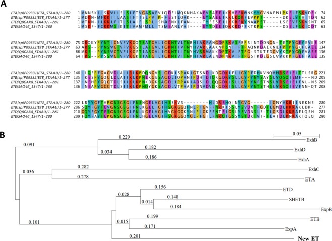
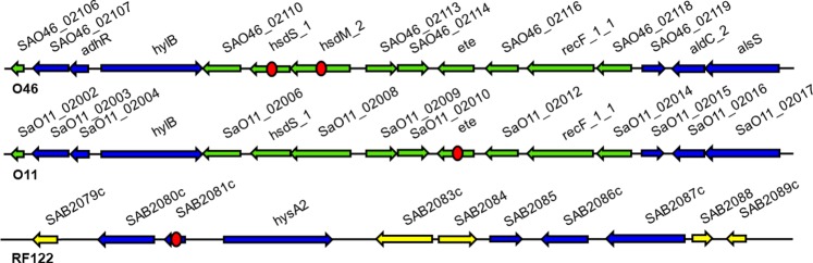
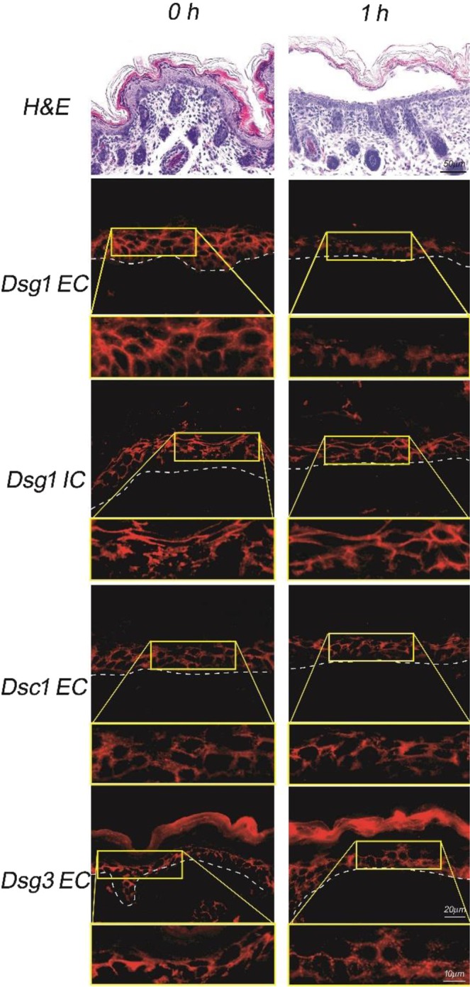
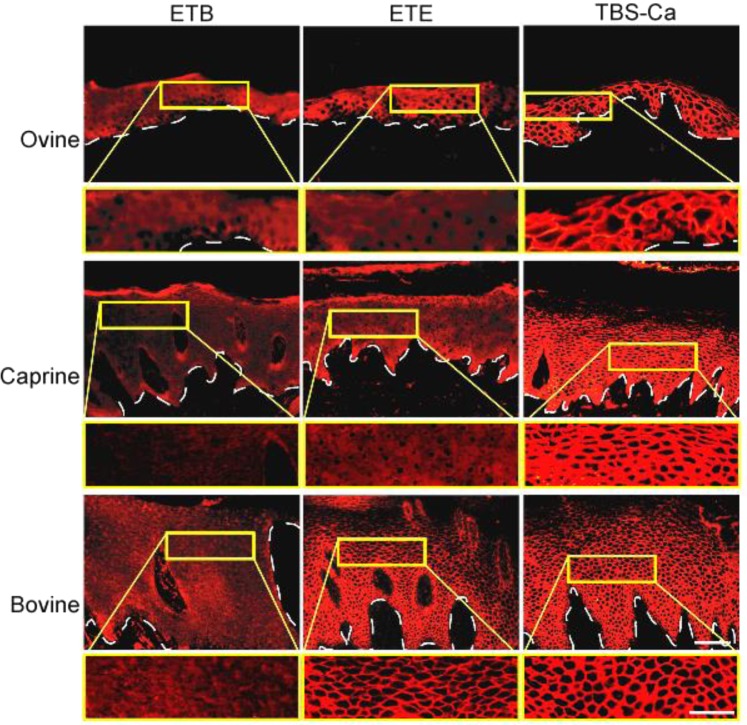
Exfoliative toxins (ETs) are secreted virulence factors produced by staphylococci. These serine proteases specifically cleave desmoglein 1 (Dsg1) in mammals and are key elements in staphylococcal skin infections. We recently identified a new et gene in S. aureus O46, a strain isolated from ovine mastitis. In the present study, we characterized the new et gene at a genetic level and the enzymatic activity of the deduced protein. The S. aureus O46 genome was re-assembled, annotated and compared with other publicly available S. aureus genomes. The deduced amino acid sequence of the new et gene shared 40%, 53% and 59% sequence identity to those of ETA, ETB and ETD, respectively. The new et gene shared the same genetic vicinity and was similar in other S. aureus strains bearing this gene. The recombinant enzyme of the new et gene caused skin exfoliation in vivo in neonatal mice. The new et-gene was thus named ete, encoding a new type (type E) of exfoliative toxin. We showed that ETE degraded the extracellular segments of Dsg1 in murine, ovine and caprine epidermis, as well as in ovine teat canal epithelia, but not that in bovine epidermis. We further showed that it directly hydrolyzed human and swine Dsg1 as well as murine Dsg1α and Dsg1β, but not canine Dsg1 or murine Dsg1γ. Molecular modeling revealed a correlation between the preferred orientation of ETE docking on its Dsg1 cleavage site and species-specific cleavage activity, suggesting that the docking step preceding cleavage accounts for the ETE species-specificity. This new virulence factor may contribute to the bacterial colonization on the stratified epithelia in certain ruminants with mastitis.


Conflict of interest statement

The authors declare no competing interests.

Figures

Figure 1
Figure 1
(A) Amino acid sequence alignment and phylogenetic tree of staphylococcal ETs. (A) Sequences retrieved from Uniprot were aligned with ClustalW with default parameters. (B) A phylogenetic analysis based on the overall amino acid sequences of ETs was built using a neighbor-joining method. SHETB, ExhA, ExhB, ExhC, and ExhD are ETs produced by Staphylococcus hyicus. ExpA and ExpB are ETs produced by Staphylococcus pseudintermedius. Three main clusters were observed, which group ExhA, ExhD and ExhB; ExhC and ETA; ExpA, ExpB, SHETB, ETB, ETD and the new ET (ETE).
Figure 2
Figure 2
Putative genomic island containing the new et gene found in the S. aureus O46 genome. Comparisons with the most closely related putative genomic islands (GI) in strains O11 and RF122, isolated from ovine and bovine hosts, respectively, are shown below the upper line. Arrows represent open reading frames and their orientations. Blue: genes shared among O46, O11, and RF122 GIs. Green: genes shared between O46 and O11 GIs. Yellow: genes only present in the RF122 GI. Red circles indicate genes lacking part of their encoding sequence (hsdM_2 in O46, ete in O11) or presenting a frameshift that results in a coding sequence truncation (hsdS_1 in O46, SAB2081c in RF122).
Figure 3
Figure 3
Exfoliative activity of the new ET in neonatal mice. Neonatal mice injected with a recombinant new ET displayed microscopic blisters 1 h after injection. Dotted lines indicate the basement membrane. Detailed views of microscopic blisters (areas delineated in yellow frame) are provided below each panel, at a higher magnification. Bars indicate 20 μm and 10 μm at lower and higher magnifications, respectively. EC: extracellular domain of Dsg, IC: intracellular domain of Dsg.
Figure 4
Figure 4
ETE degrades Dsg1 in ovine and caprine epidermis. Cryosectioned ovine, caprine and bovine nasal planum was incubated with ETB, the ETE protein or TBS-Ca, and subjected to immunofluorescence with human PF serum containing anti-Dsg1 IgG. Arrowheads indicate epidermal basement membranes. Detailed views of microscopic blisters (areas delineated in yellow frame) are provided below each panel, at a higher magnification. Bars indicate 50 μm and 20 μm at lower and higher magnifications, respectively.
Figure 5
Figure 5
ETE degrades Dsg1 in caprine teat canal epithelia. Cryosections of caprine epidermis and teat canal were incubated with either TBS-Ca or ETE, and subjected to immunofluorescence with the human PF serum. Arrowheads indicate basement membranes. Detailed views of microscopic blisters (areas delineated in yellow frame) are provided below each panel, at a higher magnification. Bars indicate 50 μm and 20 μm at lower and higher magnifications, respectively.
Figure 6
Figure 6
In vitro digestion of recombinant Dsg1s with ETE. Baculovirus recombinant extracellular domains of human Dsg1 (hDsg1), swine Dsg1 (sDsg1), canine Dsg1 (cDsg1), murine Dsg1α (mDsg1α), Dsg1β (mDsg1β) and Dsg1γ (mDsg1γ) were incubated with ETB (lane 1), ETE (lane 2), or TBS-Ca (lane 3), and subjected to immunoblotting with anti-E-tag monoclonal antibody. Original, non-cropped gel image is provided in the supplemental information file.
Figure 7
Figure 7
Protein sequence alignment and structural model of Dsg1. (A) Dsg1 protein sequence alignment of the extracellular domain. The boundaries of each cadherin repeat (EC1 to EC4) are indicated by a purple dashed line. The cleavage site is indicated by a yellow line. Residue numbers are according to model structures. (B) Superimposition of Dsg1 model structures. Ovine Dsg1 in green, human Dsg1 in blue, bovine Dsg1 in black and canine Dsg1 in red. Colored spheres are calcium ions. The cleavage site is indicated. (C) Zoom on cleavage site with the side chain of cleaved Glu332 represented and the unique calcium ion.
Figure 8
Figure 8
Best HADDOCK docking solutions of ETE on bovine and ovine Dsg1. ETE is colored from N terminus in pink to C terminus in yellow. (A) Orientation OFF (no cleavage) of ETE on bovine desmoglein (in black). (B) Orientation ON (cleavage) of ETE on ovine desmoglein (in green). For both bovine and ovine Dsg1, only EC2 to EC4 domains are shown.

Similar articles

Cited by

References

    1. Peton V, Le Loir Y. Staphylococcus aureus in veterinary medicine. Infect. Genet. Evol. 2014;21:602–615. doi: 10.1016/j.meegid.2013.08.011. - DOI - PubMed
    1. Alves PDD, et al. Molecular characterisation of Staphylococcus aureus strains isolated from small and large ruminants reveals a host rather than tissue specificity. Vet. Microbiol. 2009;137:190–195. doi: 10.1016/j.vetmic.2008.12.014. - DOI - PubMed
    1. Ben Zakour NL, et al. Genome-Wide Analysis of Ruminant Staphylococcus aureus Reveals Diversification of the Core Genome. J. Bacteriol. 2008;190:6302–6317. doi: 10.1128/JB.01984-07. - DOI - PMC - PubMed
    1. Guinane CM, et al. Evolutionary Genomics of Staphylococcus aureus Reveals Insights into the Origin and Molecular Basis of Ruminant Host Adaptation. Genome Biol. Evol. 2010;2:454–466. doi: 10.1093/gbe/evq031. - DOI - PMC - PubMed
    1. Nishifuji K, Sugai M, Amagai M. Staphylococcal exfoliative toxins: “Molecular scissors” of bacteria that attack the cutaneous defense barrier in mammals. J. Dermatol. Sci. 2008;49:21–31. doi: 10.1016/j.jdermsci.2007.05.007. - DOI - PubMed

Publication types

Substances

LinkOut - more resources