. 2020 Jan;196(1):85-94.
doi: 10.1007/s00066-019-01532-8. Epub 2019 Nov 8.

Modulation of radiation-induced oral mucositis (mouse) by dermatan sulfate: effects on differentiation processes

Affiliations

Modulation of radiation-induced oral mucositis (mouse) by dermatan sulfate: effects on differentiation processes

Nilsu Cini et al. Strahlenther Onkol. 2020 Jan.

Abstract

Purpose: During head and neck cancer radiotherapy, oral mucositis is the most frequent early side effect. Systemic dermatan sulfate (DS) administration has been shown to significantly decrease oral mucosal radiation reactions during daily fractionated irradiation (IR) in an established mouse model. The aim of this study was to investigate the mechanism of the oral epithelial differentiation process, during IR alone and in combination with DS treatment in the same mouse model.

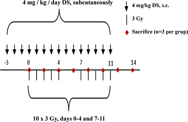
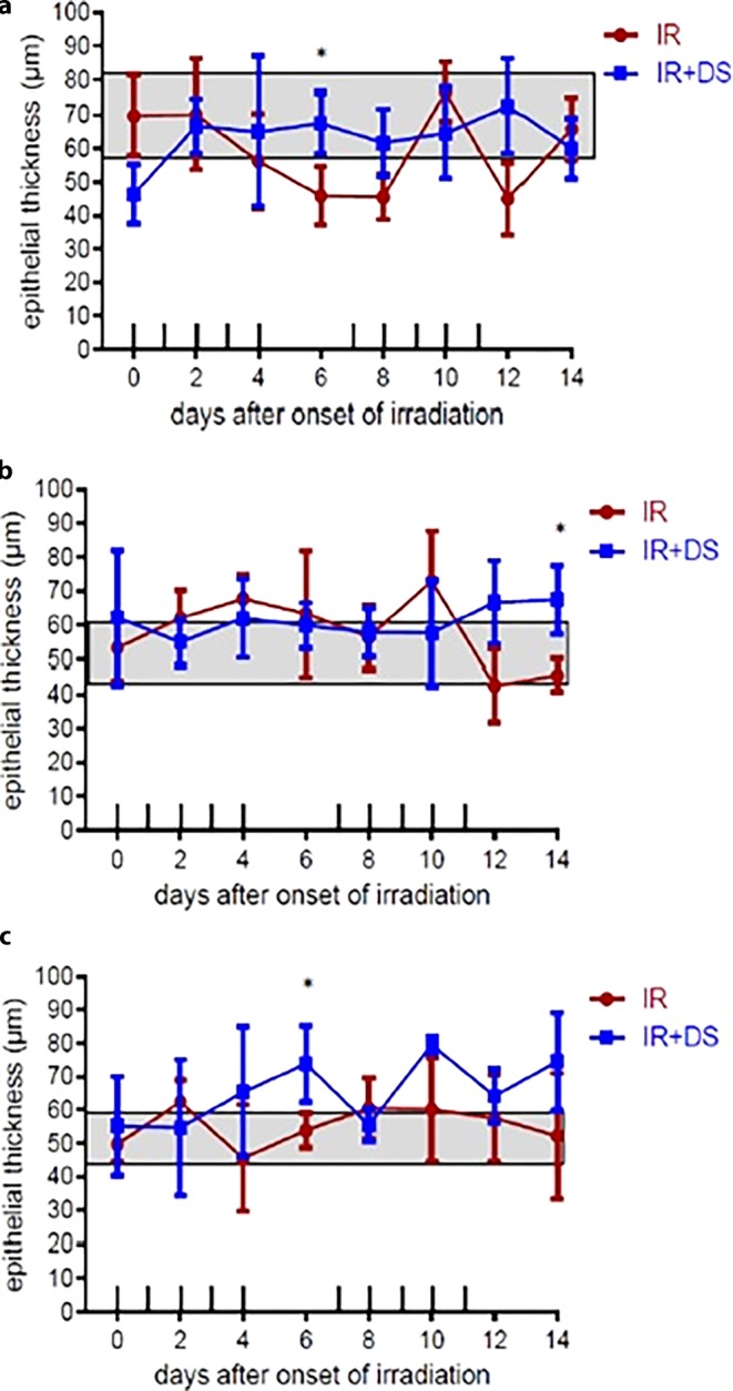
Methods: Fractionated IR 5 × 3 Gy/week was given to the snouts of mice over two weeks, either alone (IR) or in combination with daily DS treatment of 4 mg/kg (IR + DS). Groups of mice (n = 3) were sacrificed every second day over the course of 14 days in both experimental arms. Their tongue was excised and subjected to immunohistochemical processing.

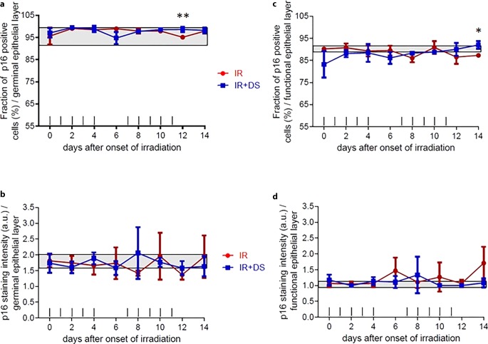
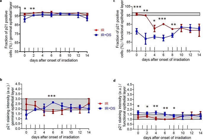
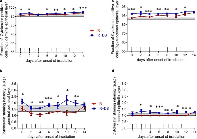
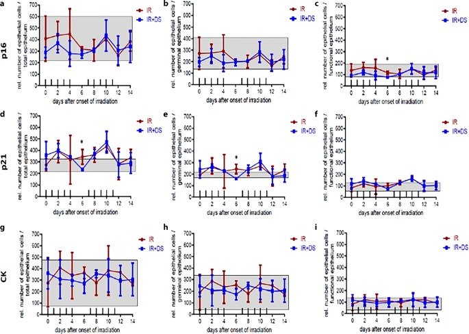
Results: In the p16 analysis as a proliferation marker, the difference between IR alone and IR + DS in the germinal (proliferation) layer was not significant, not stimulating the proliferation process. For the p21 analysis as a differentiation marker on the functional (differentiation) layer, the difference between IR alone and IR + DS arms was significant, indicating that DS inhibited the differentiation process. In the cytokeratin (CK) analysis as the indicator of cellular skeletal integrity, the percentage of antibody-positive cells was above the normal level in both experimental arms and significantly superior in the IR + DS arm.

Conclusion: The mucosal protective activity of DS, instead of stimulating proliferation, is based on prevention of cell loss by a combination of effects leading to the inhibition of cellular differentiation and an increase in the expression of epithelial mechanical strength between intercellular mechanical junctions.

Keywords: Cellular junctions; Differentiation; Fractionation; Mechanical strength; Proliferation.


Conflict of interest statement

N. Cini, S. Gruber, Z. Arican Alicikus, and W. Dörr declare that they have no competing interests.

Figures

Fig. 1
Fig. 1
Experimental design. IR comprised fractionated IR with 3 Gy per day over two weeks. No IR was given at the weekend (days 5–6 and 12–13). IR was applied either alone or in combination with daily 4 mg/kg DS treatment over 14 days. At two-day intervals, 3 animals were sacrificed per experimental group, 3 untreated mice (day 0) served as controls
Fig. 2
Fig. 2
p16, p21, and CK expression during fractionated IR ± DS. Representative histophotographs of p16-, p21-, and CK-stained lower mouse tongue epithelia were taken on day 0 (unirradiated and untreated controls) and day 14. Scale bar: 50 µm
Fig. 3
Fig. 3
Effect of fractionated IR ± DS number of cells immunohistochemically positive stained cell percentage and staining intensity for p16. p16 expression changes were analyzed in the germinal and the functional epithelial compartment. The staining signal intensity was scored semi-quantitatively with an arbitrary score of 0 (no signal), 1 (weak), 2 (intermediate), or a maximum of 3 (strong). p16 was analyzed in 3 specimens per experimental arm, every second day over the course of 14 days. Data points represent the mean of 3 animals, error bars indicate ±1 SEM. The shaded areas illustrate the mean (±1 SEM) from 3 control animals. The fractionation protocol is indicated on top of the abscissae. Asteriskp < 0.05; doubleAsteriskp < 0.01
Fig. 4
Fig. 4
Effect of fractionated IR ± DS number of cells immunohistochemically positive stained cell percentage and staining intensity for p21. p21 expression changes were analyzed in the germinal and the functional epithelial compartment. The staining signal intensity was scored semi-quantitatively with an arbitrary score of 0 (no signal), 1 (weak), 2 (intermediate), or a maximum of 3 (strong). p21 was analyzed in 3 specimens per experimental arm, every second day over the course of 14 days. Data points represent the mean of 3 animals, error bars indicate ±1 SEM. The shaded areas illustrate the mean (±1 SEM) from 3 control animals. The fractionation protocol is indicated on top of the abscissae. Asteriskp < 0.05; double Asteriskp < 0.01; tripleAsteriskp < 0.001
Fig. 5
Fig. 5
Effect of fractionated IR ± DS number of cells immunohistochemically positive stained cell percentage and staining intensity for CK. CK expression changes were analyzed in the germinal and the functional epithelial compartment. The staining signal intensity was scored semi-quantitatively with an arbitrary score of 0 (no signal), 1 (weak), 2 (intermediate), or a maximum of 3 (strong). CK was analyzed in 3 specimens per experimental arm, every second day over the course of 14 days. Data points represent the mean of 3 animals, error bars indicate ±1 SEM. The shaded areas illustrate the mean (±1 SEM) from 3 control animals. The fractionation protocol is indicated on top of the abscissae. Asteriskp < 0.05; double Asteriskp < 0.01; tripleAsteriskp < 0.001
Fig. 6
Fig. 6
Relative epithelial thickness during daily fractionated IR ± DS treatment on the epithelial expression of p16, p21, and CK. Epithelial thickness was determined at representative spots in each tongue section. p16, p21, and CK were analyzed in 3 specimens per experimental arm, every second day over the course of 14 days. Data points represent the mean of 3 animals, error bars indicate ±1 SEM. The shaded areas illustrate the mean (±1 SEM) from 3 control animals. The fractionation protocol is indicated on top of the abscissae. Asteriskp < 0.05; doubleAsteriskp < 0.01; tripleAsteriskp < 0.001
Fig. 7
Fig. 7
Relative number of epithelial cells during daily fractionated IR ± DS treatment on the epithelial expression of p16, p21, and CK for total epithelium, germinal epithelium, and functional epithelium. Cell numbers were determined at representative spots in each tongue section. p16, p21, and CK were analyzed in 3 specimens per experimental arm, every second day over the course of 14 days. Data points represent the mean of 3 animals, error bars indicate ±1 SEM. The shaded areas illustrate the mean (±1 SEM) from 3 control animals. The fractionation protocol is indicated on top of the abscissae. Asteriskp < 0.05; doubleAsteriskp < 0.01; tripleAsteriskp < 0.001

References

    1. Sonis ST. Oral mucosits in head and neck cancer: risk, biology, and management. Am Soc Clin Oncol Educ Book. 2013;33:236–240. - PubMed
    1. Rodríguez-Caballero A, Torres-Lagares D, Robles-García M, et al. Cancer treatment-induced oral mucositis: a critical review. Int J Oral Maxillofac Surg. 2012;41(2):225–238. - PubMed
    1. Agbaje JO, Jacobs R, Michiels K, et al. Bone healing after dental extractions in irradiated patients: a pilot study on a novel technique for volume assessment of healing tooth sockets. Clin Oral Investig. 2009;13(3):257–261. - PubMed
    1. Squier CA, Kremer MJ. Biology of oral mucosa and esophagus. J Natl Cancer Inst Monogr. 2001;29:7–15. - PubMed
    1. Qin R, Steel A, Fazel N. Oral mucosa biology and salivary biomarkers. Clin Dermatol. 2017;35(5):477–483. - PubMed

MeSH terms