. 2020 Feb;249(2):245-261.
doi: 10.1002/dvdy.130. Epub 2019 Nov 20.

Zebrafish etv2 knock-in line labels vascular endothelial and blood progenitor cells

Affiliations

Zebrafish etv2 knock-in line labels vascular endothelial and blood progenitor cells

Brendan Chestnut et al. Dev Dyn. 2020 Feb.

Abstract

Background: ETS transcription factor Etv2/Etsrp is one of the earliest markers for vascular and hematopoietic progenitors and functions as a key regulator of hematovascular development in multiple vertebrates, including zebrafish. Therefore, transgenic etv2 reporter lines provide a valuable tool to study vasculogenesis and hematopoiesis. However, previously generated zebrafish reporter lines do not fully recapitulate the endogenous pattern of etv2 expression.

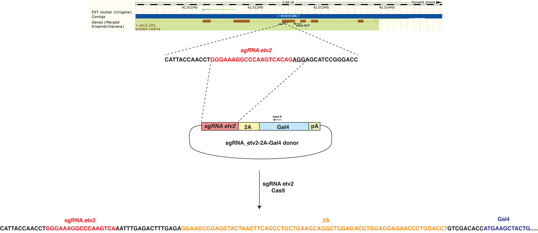
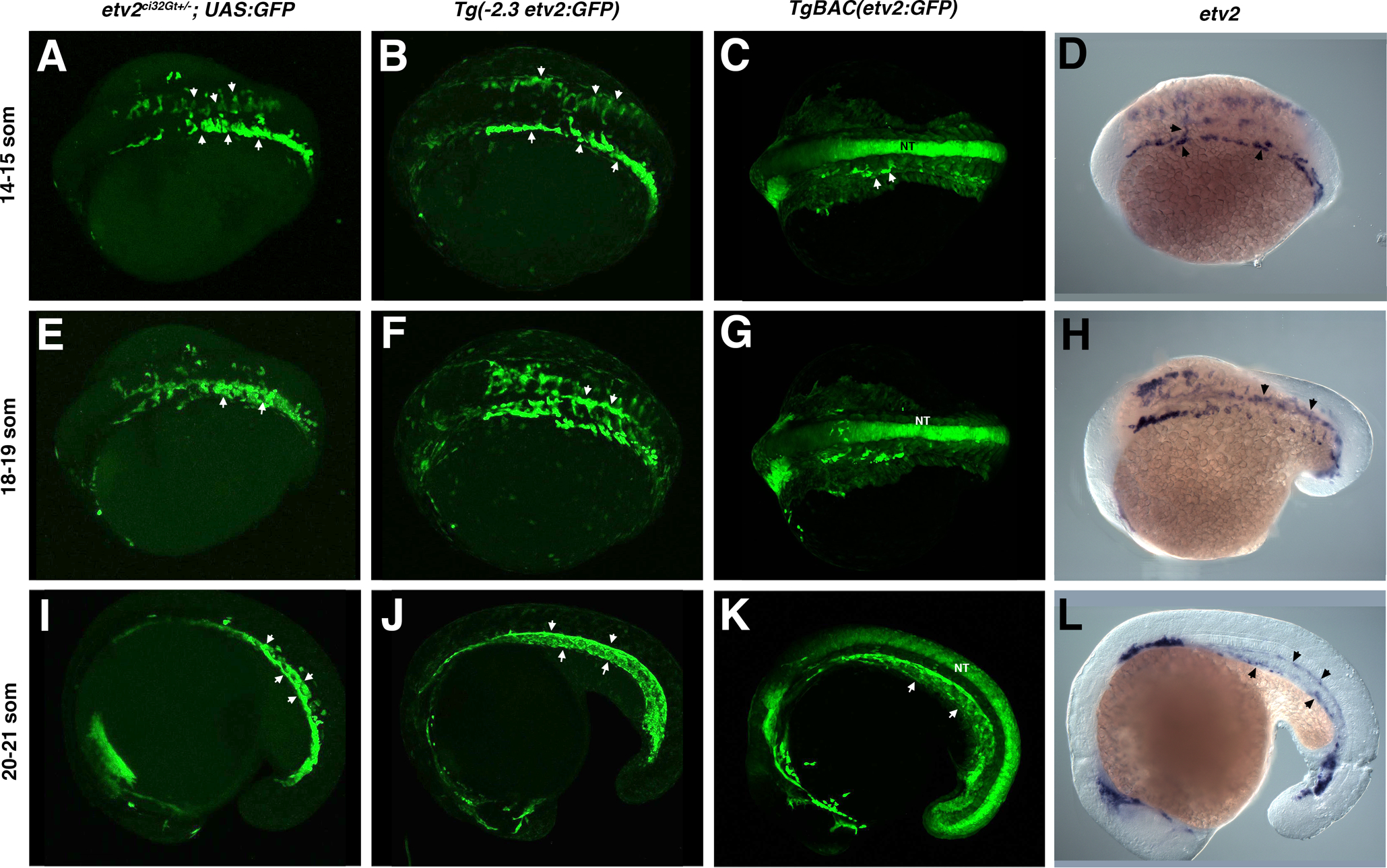
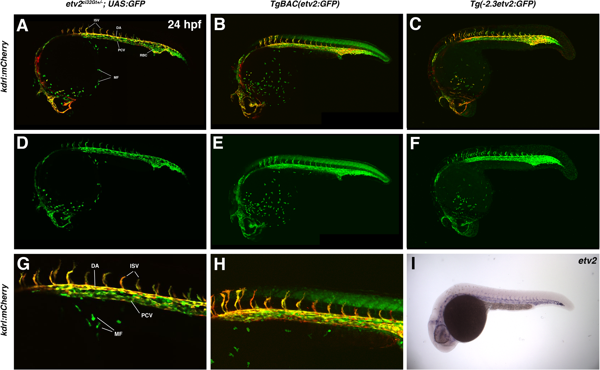
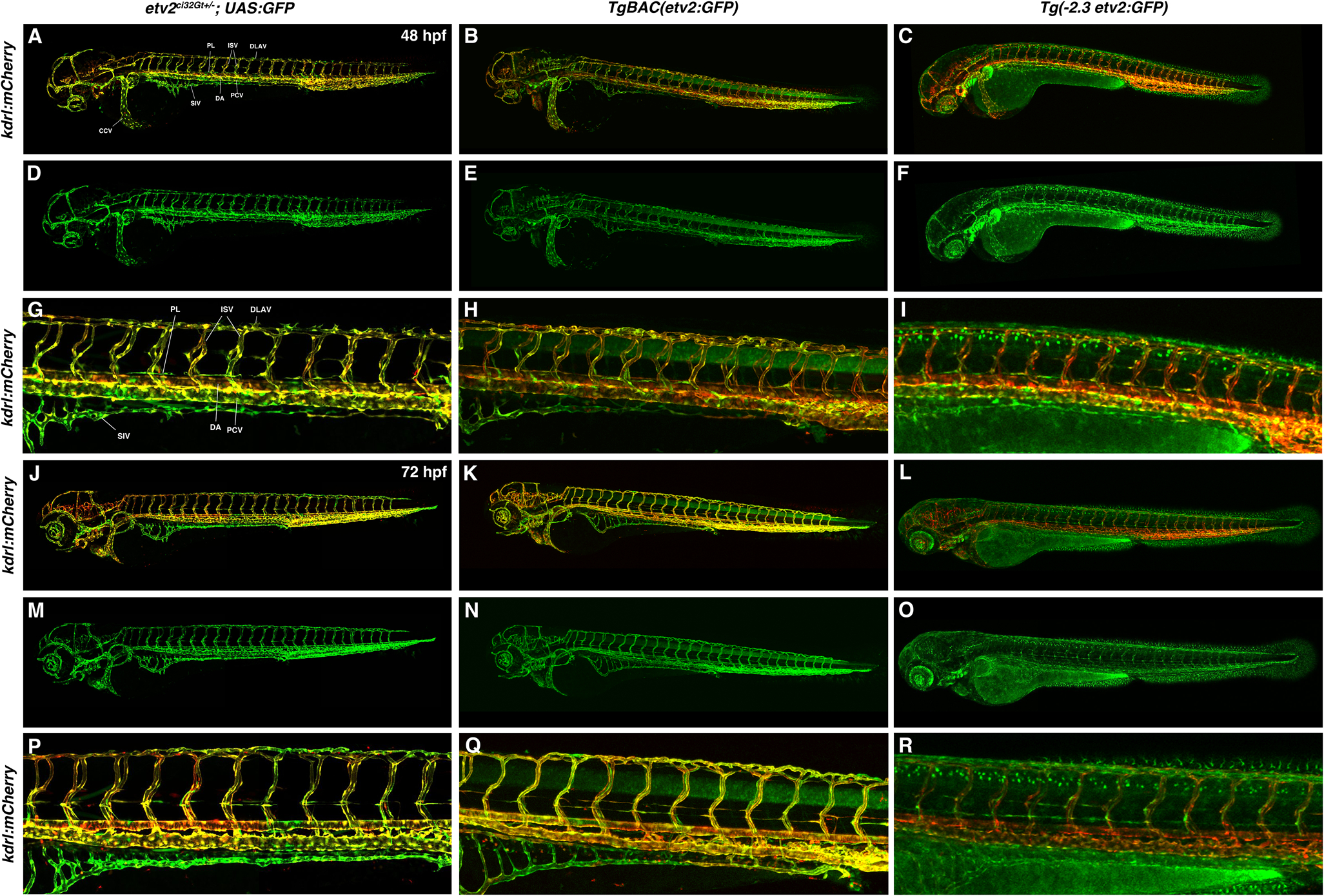
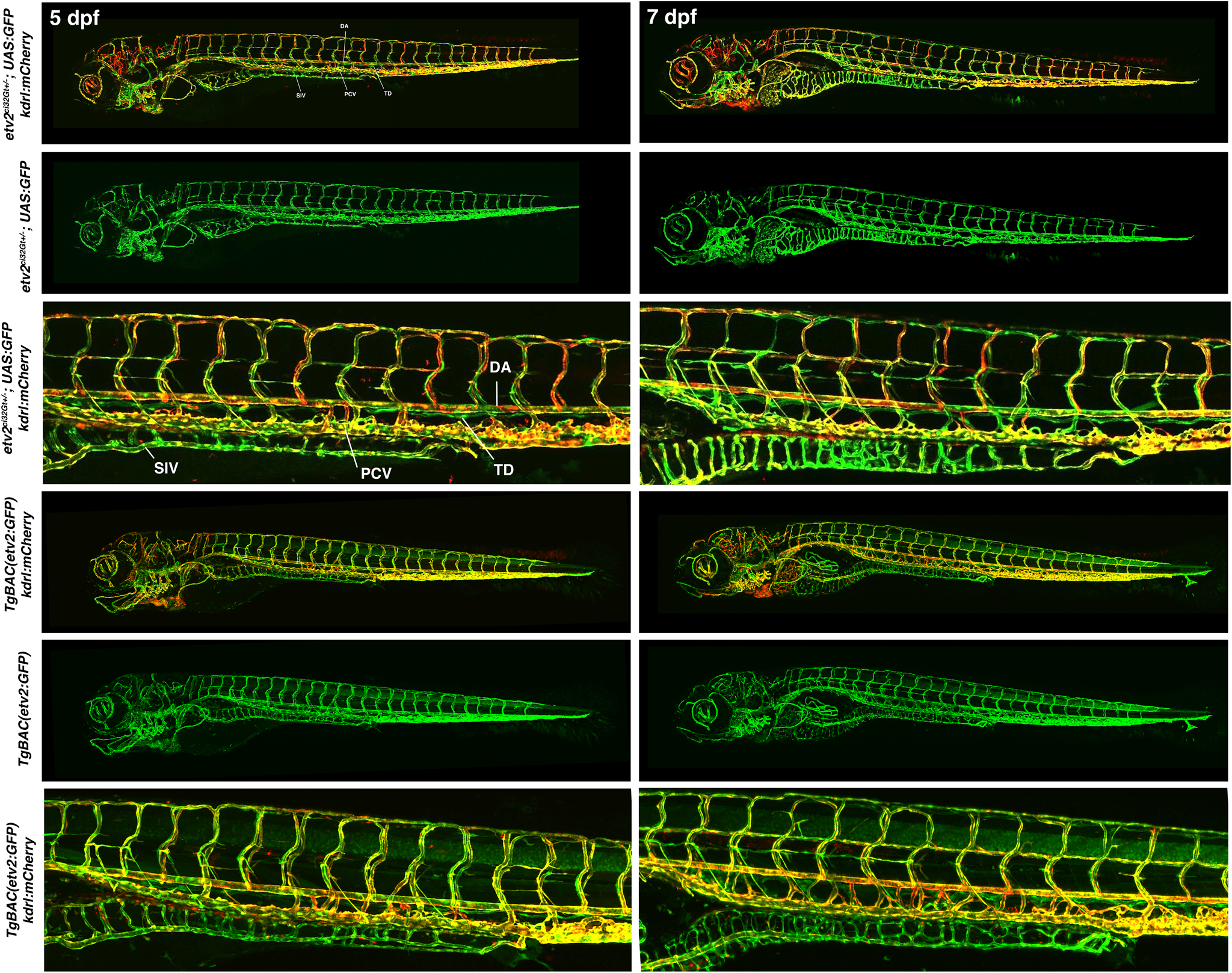
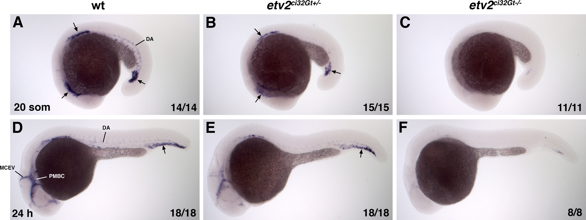
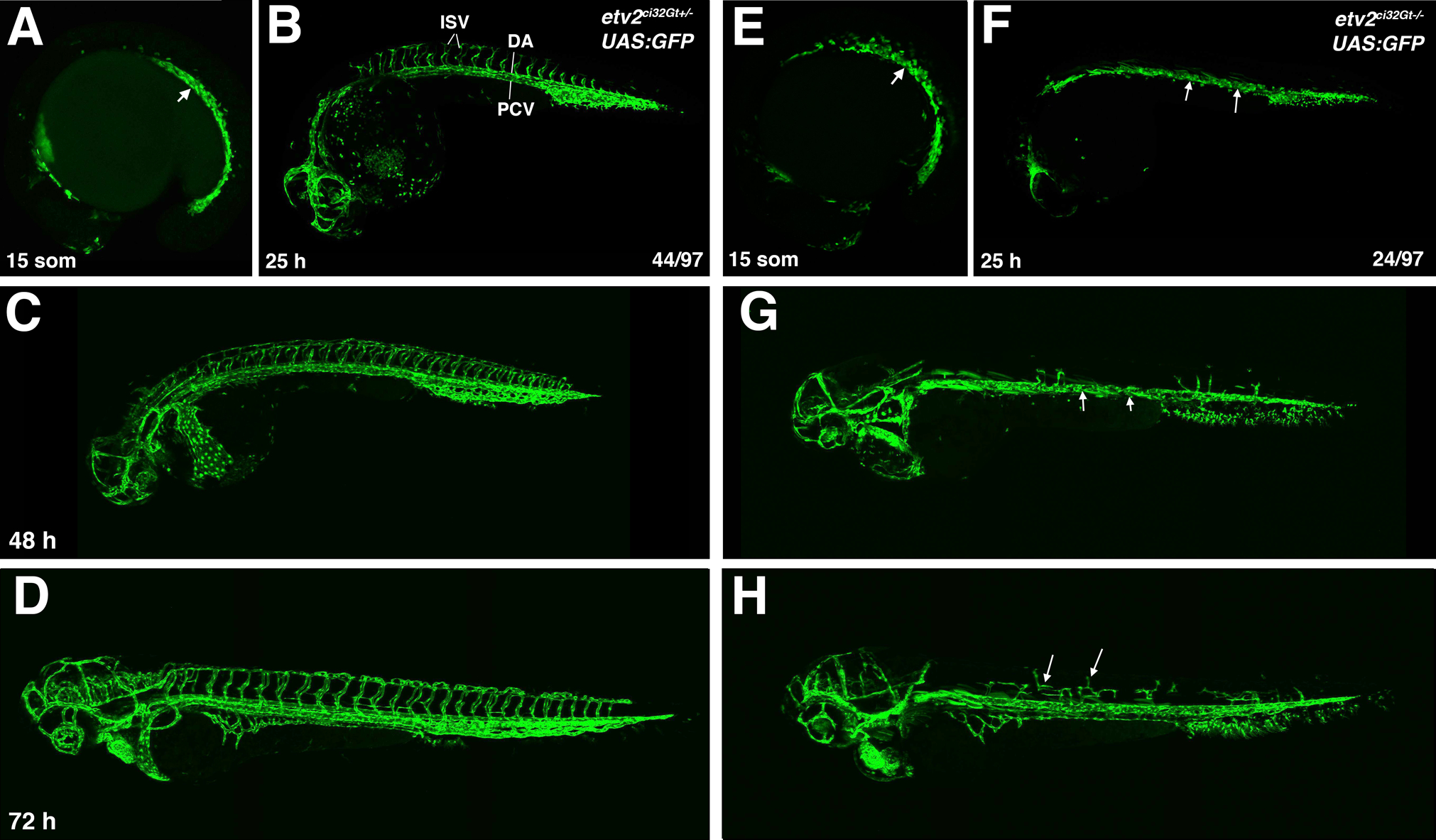
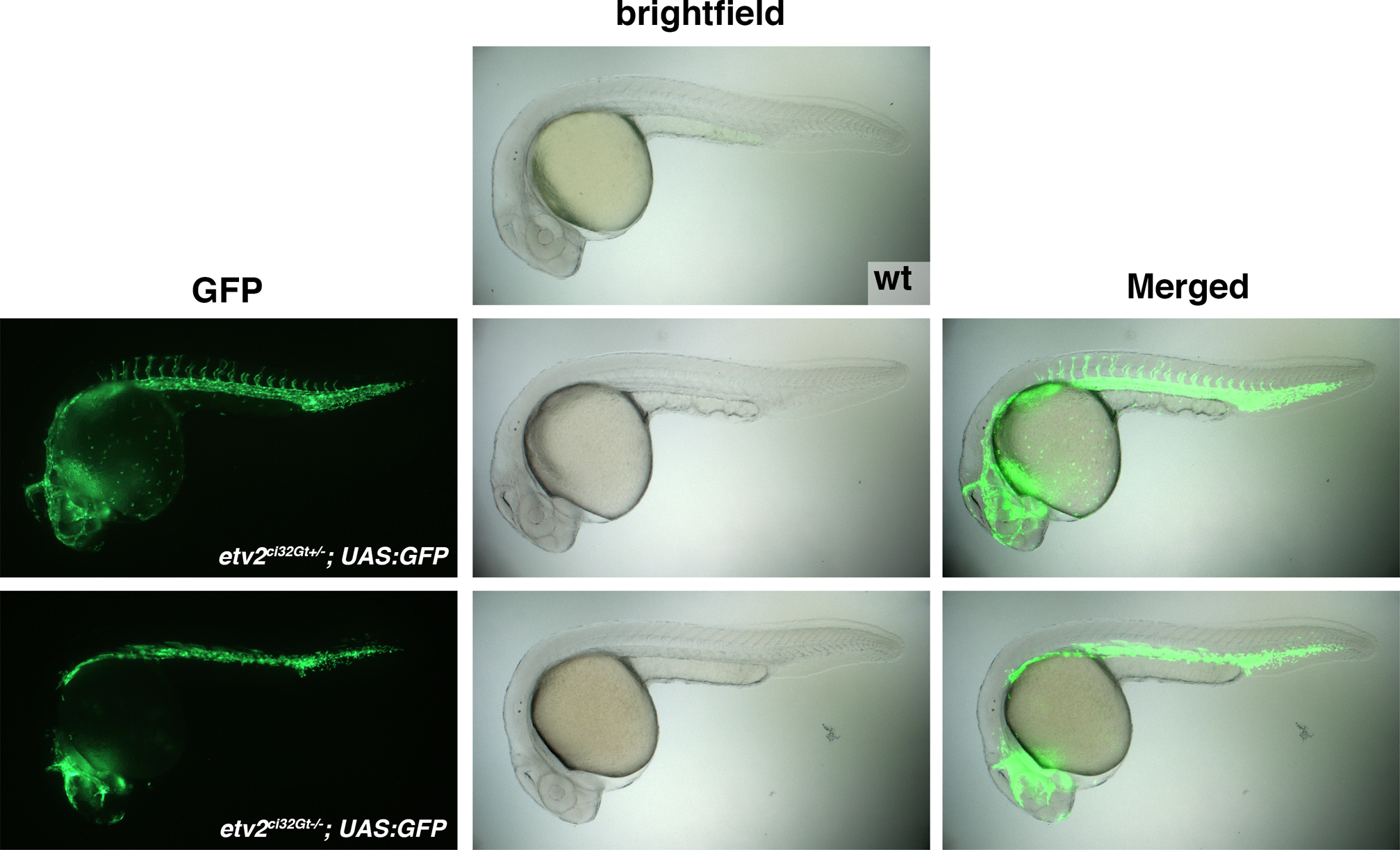
Results: Here we used CRISPR/Cas9-mediated homology-independent DNA repair approach to knock-in a Gal4 transcriptional activator into the zebrafish etv2 genomic locus, thus generating etv2 ci32Gt gene trap line. etv2 ci32Gt ; UAS:GFP embryos show GFP expression in vascular endothelial, myeloid and red blood cells. Because gal4 insertion interrupts the etv2 locus, homozygous etv2 ci32Gt embryos display defects in vasculogenesis and myelopoiesis, and enable visualizing etv2-deficient hematovascular progenitors in live embryos. Furthermore, we performed differential transcriptome analysis of sorted GFP-positive cells from heterozygous and homozygous etv2 ci32Gt embryos. Approximately 500 downregulated genes were identified in etv2 ci32Gt homozygous embryos, which include multiple genes expressed in vascular endothelial and myeloid cells.

Conclusions: The etv2 ci32Gt gene trap line and the data sets of misregulated genes will be valuable resources to study hematopoietic and vascular development.

Keywords: CRISPR; Cas9; RNA-seq; myeloid; red blood cell; transcriptome; vascular endothelial; zebrafish.


Figures

Figure 1.
Figure 1.
A diagram of sgRNA-2A-Gal4 construct and its insertion into the etv2 genomic locus. The targeting construct contained etv2 sgRNA site, followed by in-frame fusion to the viral peptide P2A, a transcriptional activator Gal4 and the SV40 polyA sequence. etv2 sgRNA targets the fifth exon of etv2 genomic sequence. The diagram shows approximate locations (not to scale) of the primers used for PCR amplification and sequencing.
Figure 2.
Figure 2.
Effect of different mutations on expression of the etv2-2A-GFP reporter. The reporter was designed to mimic the insertion of 2A-Gal4 into the genomic etv2 locus. Wild-type (wt) construct contains 5’UTR and the N-terminal coding sequence of the etv2 gene, and 2A-GFP sequence inserted out-of-frame. Four potential translation-initiating ATG sequences are shown in Frame 1. Frame 2 contains a single initiating ATG which is in-frame with 2A-GFP translation. Mutant 1 (Mut1) has mutations in all four ATG sites present in frame 1. Mutant 2 (Mut2) has mutation in the ATG site within the frame 2. Mutant 3 (Mut3) has a mutation within the ATG sequence that initiates GFP translation. The scatter plot shows relative GFP fluorescence intensity values (plotted in log2 scale) of embryos injected with different etv2–2A-GFP reporter constructs. Note increased GFP fluorescence in embryos injected with Mut1 construct and reduced expression in Mut2 and Mut3-injected embryos compared to embryos injected with the wt construct. ** p<0.01, *** p<0.001, ****p<0.0001, t-Student’s test (two-tailed).
Figure 3.
Figure 3.
A comparison of etv2ci32Gt+/−; UAS:GFP, Tg(−2.3etv2:GFP), TgBAC(etv2:GFP) fluorescence pattern and etv2 mRNA expression analyzed by in situ hybridization (ISH) at the 14–21 somite stages. (A-C) GFP fluorescence or (D) etv2 mRNA expression is apparent in bilaterally located vascular endothelial progenitors which have started migrating towards the midline at the 14–15-somite stages (arrowheads). Strong non-specific GFP expression in the neural tube (NT) is apparent in TgBAC(etv2:GFP) embryos. (E-H) GFP-positive vascular endothelial progenitors at the 18–19-somite stages are coalescing at the midline into vascular cords (arrowheads, E,F). Similar etv2 expression pattern is observed by ISH analysis (H). (I-L) GFP expression at the 20–21-somite stages is apparent in the forming axial vasculature (arrowheads), and non-specific expression is apparent in the neural tube (K). Similar etv2 expression pattern is apparent from ISH analysis (L). A-H, dorsal or dorso-lateral view; I-L, lateral view, anterior is to the left.
Figure 4.
Figure 4.
A comparison of etv2ci32Gt+/−; UAS:GFP, TgBAC(etv2:GFP), Tg(−2.3etv2:GFP) fluorescence pattern (in kdrl:mCherry background, top row) and etv2 mRNA expression analyzed by in situ hybridization (ISH) at 24 hpf. etv2ci32Gt+/−; UAS:GFP expression is apparent throughout the entire vasculature, red blood cells (RBC) and macrophages (MF). TgBAC(etv2:GFP) shows similar expression pattern, and also shows non-specific expression in the neural tube. (A-C) Merged mCherry and GFP channels; (D-F) GFP channel; (G,H) magnified images of the trunk region in (A,B). (I) ISH analysis for etv2 mRNA expression at 24 hpf. DA, dorsal aorta; PCV, posterior cardinal vein; ISV, intersegmental vessels.
Figure 5.
Figure 5.
A comparison of etv2ci32Gt+/−; UAS:GFP, TgBAC(etv2:GFP), Tg(−2.3etv2:GFP) fluorescence pattern (in kdrl:mCherry background) at 48 hpf (A-I) and 72 hpf (J-R). etv2ci32Gt+/−; UAS:GFP expression is apparent throughout the entire vasculature and in lymphatic progenitors (parachordal lymphangioblasts, PLs). TgBAC(etv2:GFP) shows similar expression pattern, and also shows non-specific expression in the neural tube. Vascular endothelial expression in Tg(−2.3etv2:GFP) line is downregulated after 48 hpf and is very weak at 72 hpf, while non-specific epithelial expression is apparent. (A-C, J-L) Merged mCherry and GFP channels; (D-F, M-O) GFP channel; (G-I, P-R) magnified images of the trunk region in (A-C, J-L). DA, dorsal aorta; PCV, posterior cardinal vein; ISV, intersegmental vessels, SIV, subintestinal vessel; DLAV, dorsal longitudinal anastomotic vessel; CCV, common cardinal vein.
Figure 6.
Figure 6.
A comparison of etv2ci32Gt+/−; UAS:GFP and TgBAC(etv2:GFP) embryos in kdrl:mCherry background at 5 and 7 dpf. Both lines show GFP expression in the entire vasculature and lymphatics. DA, dorsal aorta; PCV, posterior cardinal vein; SIV, subintestinal vein (thoracic duct, TD). Tg (−2.3etv2:GFP) line did not show vascular endothelial expression at these stages.
Figure 7.
Figure 7.
etv2 expression analysis in wild-type, etv2ci32G+−/− and etv2ci32Gt−/− embryos at the 20-somite (A-C) and 24 hpf stages (D-F). Embryos were obtained from an incross of etv2ci32Gt+/−; UAS:GFP+/+ parents and sorted based on their GFP fluorescence pattern. Antisense etv2 RNA probe which corresponds to the C-terminal portion of the etv2 coding sequence and 3’UTR downstream of the Gal4 insertion side was used for in situ hybridization (see Experimental Procedures). (A-C) In non-fluorescent wild-type siblings (wt), strong etv2 expression is apparent in vascular progenitors in the anterior lateral plate mesoderm, presumptive progenitors of the anterior and common cardinal veins, and in vascular progenitors next to the tailbud (arrows). Weaker expression in the dorsal aorta (DA) is also apparent. Note the reduced etv2 expression in etv2ci32Gt+/− embryos and nearly absent expression in etv2ci32Gt−/− embryos. (D-F) In wild-type siblings, strong etv2 expression is apparent in the cranial venous vasculature, including the primordial midbrain channel (PMBC) and the middle cerebral vein (MCEV), as well as the tail plexus region (arrow). Weaker expression in the DA is also apparent. Note the reduced etv2 expression in etv2ci32Gt+/− embryos and nearly absent expression in etv2ci32Gt−/− embryos.
Figure 8.
Figure 8.
A comparison of vascular development in heterozygous etv2ci32Gt+/−; UAS:GFP and homozygous etv2ci32Gt−/−; UAS:GFP embryos. GFP expression is apparent in vascular progenitors of the trunk axial vasculature (arrow, A) in etv2ci32Gt+/−; UAS:GFP embryos. This domain is broader and disorganized in etv2ci32Gt−/−; UAS:GFP embryos (E). Vascular progenitors fail to coalesce into vascular cords in etv2ci32Gt−/−; UAS:GFP embryos at 25 hpf (arrows, F, compare with C). Intersegmental vessels (ISVs) are absent in etv2ci32Gt−/−; UAS:GFP embryos between 25–48 hpf (F,G). Some ISV sprouts are apparent at 72 hpf but are shorter and mispatterned (H, arrows).
Figure 9.
Figure 9.
Brightfield and fluorescent images of etv2ci32Gt+/− and etv2ci32Gt−/− embryos and their non-fluorescent wild-type siblings at 24 hpf. No morphological defects are apparent in the brightfield images of etv2ci32Gt+/− and etv2ci32Gt−/− embryos.

References

    1. Asakawa K, Suster ML, Mizusawa K, Nagayoshi S, Kotani T, Urasaki A, Kishimoto Y, Hibi M, Kawakami K. 2008. Genetic dissection of neural circuits by Tol2 transposon-mediated Gal4 gene and enhancer trapping in zebrafish. Proc Natl Acad Sci U S A 105:1255–1260. - PMC - PubMed
    1. Auer TO, Duroure K, De Cian A, Concordet JP, Del Bene F. 2014. Highly efficient CRISPR/Cas9-mediated knock-in in zebrafish by homology-independent DNA repair. Genome Res 24:142–153. - PMC - PubMed
    1. Balciunas D, Wangensteen KJ, Wilber A, Bell J, Geurts A, Sivasubbu S, Wang X, Hackett PB, Largaespada DA, McIvor RS, Ekker SC. 2006. Harnessing a high cargo-capacity transposon for genetic applications in vertebrates. PLoS Genet 2:e169. - PMC - PubMed
    1. Craig MP, Grajevskaja V, Liao HK, Balciuniene J, Ekker SC, Park JS, Essner JJ, Balciunas D, Sumanas S. 2015. Etv2 and fli1b function together as key regulators of vasculogenesis and angiogenesis. Arterioscler Thromb Vasc Biol 35:865–876. - PMC - PubMed
    1. Davis JA, Koenig AL, Lubert A, Chestnut B, Liu F, Palencia Desai S, Winkler T, Pociute K, Choi K, Sumanas S. 2018. ETS transcription factor Etsrp / Etv2 is required for lymphangiogenesis and directly regulates vegfr3 / flt4 expression. Dev Biol 440:40–52. - PMC - PubMed

Publication types

MeSH terms