. 2020 Jan:173:104646.
doi: 10.1016/j.antiviral.2019.104646. Epub 2019 Nov 6.

Non-invasive bioluminescence imaging of HCoV-OC43 infection and therapy in the central nervous system of live mice

Affiliations

Non-invasive bioluminescence imaging of HCoV-OC43 infection and therapy in the central nervous system of live mice

Junwei Niu et al. Antiviral Res. 2020 Jan.

Abstract

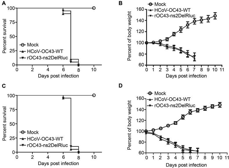
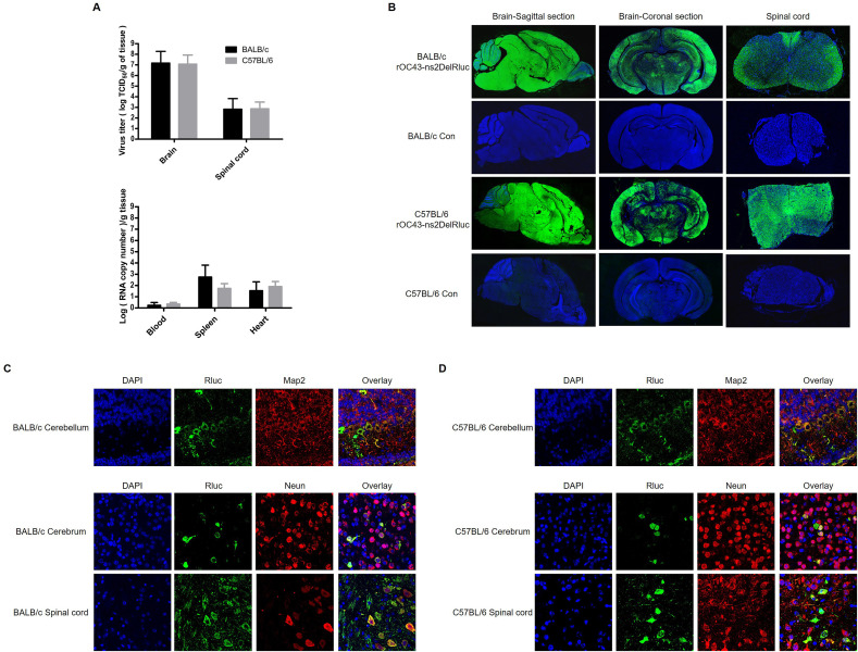
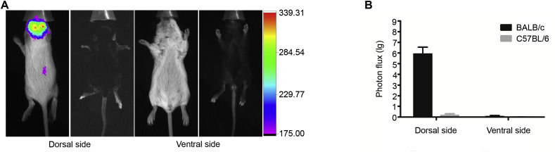
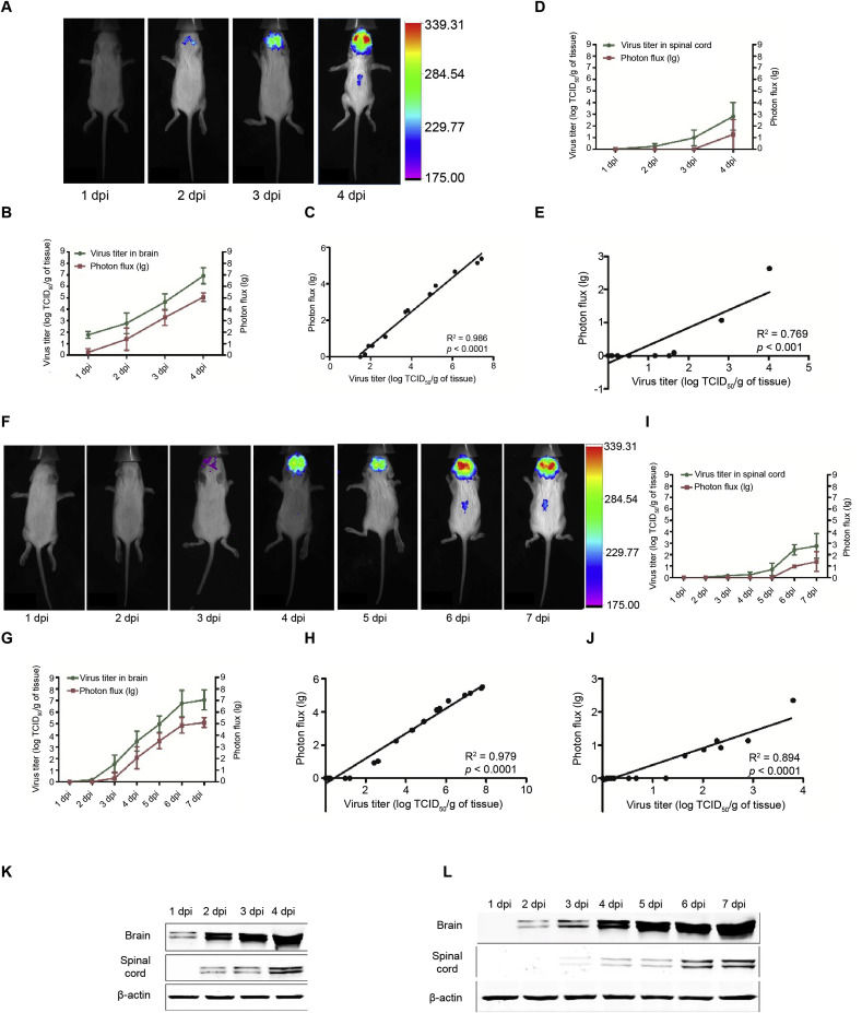
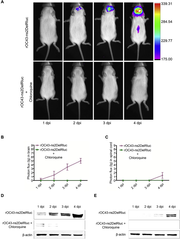
Human coronaviruses (HCoVs) are important pathogens that cause upper respiratory tract infections and have neuroinvasive abilities; however, little is known about the dynamic infection process of CoVs in vivo, and there are currently no specific antiviral drugs to prevent or treat HCoV infection. Here, we verified the replication ability and pathogenicity of a reporter HCoV-OC43 strain expressing Renilla luciferase (Rluc; rOC43-ns2DelRluc) in mice with different genetic backgrounds (C57BL/6 and BALB/c). Additionally, we monitored the spatial and temporal progression of HCoV-OC43 through the central nervous system (CNS) of live BALB/c mice after intranasal or intracerebral inoculation with rOC43-ns2DelRluc. We found that rOC43-ns2DelRluc was fatal to suckling mice after intranasal inoculation, and that viral titers and Rluc expression were detected in the brains and spinal cords of mice infected with rOC43-ns2DelRluc. Moreover, viral replication was initially observed in the brain by non-invasive bioluminescence imaging before the infection spread to the spinal cord of BALB/c mice, consistent with its tropism in the CNS. Furthermore, the Rluc readout correlated with the HCoV replication ability and protein expression, which allowed quantification of antiviral activity in live mice. Additionally, we validated that chloroquine strongly inhibited rOC43-ns2DelRluc replication in vivo. These results provide new insights into the temporal and spatial dissemination of HCoV-OC43 in the CNS, and our methods provide an extremely sensitive platform for evaluating the efficacy of antiviral therapies to treat neuroinvasive HCoVs in live mice.

Keywords: Bioluminescence imaging; Chloroquine; Coronavirus; Mouse; Therapy.


Conflict of interest statement

The authors declare no commercial or financial conflicts of interest.

Figures

Fig. 1
Fig. 1
rOC43-ns2DelRluc is fatal in suckling mice. (A and B) BALB/c mice and (C and D) C57BL/6 mice were intranasally infected with 104 TCID50 of rOC43-ns2DelRluc (n = 20/group). Weight loss (B and D) and survival (A and C) were monitored daily.
Fig. 2
Fig. 2
Replication and dissemination of rOC43-ns2DelRluc in mice with different genetic backgrounds. (A) Detection of rOC43-ns2DelRluc in different tissues. Mice were inoculated intranasally with 104 TCID50 rOC43-ns2DelRluc and sacrificed at 6 dpi. Tissue supernatants were analyzed for viral load by IFA (upper) in BHK-21 cells or real-time qRT-PCR (lower). Data represent three independent experiments and are shown as the mean ± standard deviation. (B) Immunofluorescence staining of infected and mock-infected mouse brains and spinal cords. (C and D) Double-staining for Rluc and neuron cells in brain and spinal cord sections. Mice were inoculated intranasally with 104 TCID50 rOC43-ns2DelRluc and sacrificed at 6 dpi. Brain and spinal cord tissues were analyzed using Operetta (PerkinElmer) with Columbus software (B) or a Leica TCS SP8 confocal microscope with LAS software (C and D). Representative graphs are shown of various brain and spinal cord sections from rOC43-ns2DelRluc-infected BALB/c and C57BL/6 mice.
Fig. 3
Fig. 3
BLI of rOC43-ns2DelRluc in mice with different genetic backgrounds. (A) BLI of the dorsal and ventral sides of mice with different genetic backgrounds. BALB/c and C57BL/6 mice were intranasally infected with 104 TCID50 rOC43-ns2DelRluc, and BLI was performed at 6 dpi by intraperitoneal injection of Rluc substrate and capture of photon emission using an In Vivo F Pro system. Representative graphs are shown of the dorsal and ventral views. (B) Comparison of BLI from different sides of mice with different genetic backgrounds. Data represent three independent experiments and are shown as the mean ± standard deviation.
Fig. 4
Fig. 4
BLI of the spatial and temporal progression of rOC43-ns2DelRluc infection. BLI of BALB/c mice after intracerebral (A) or intranasal (F) infection with rOC43-ns2DelRluc. BALB/c mice underwent intracerebral (100 TCID50) or intranasal (104 TCID50) inoculation on day 0 with rOC43-ns2DelRluc, and BLI was performed daily by intraperitoneal injection of Rluc substrate and capture of photon emission using an In Vivo F Pro system. Representative BLI images are shown. (B–E) Data correspond to the time course after intracerebral infection. Luciferase activity was quantified using the In Vivo F Pro system, and viral titer was determined by indirect IFA of brain (B) and spinal cord (D) tissue at the indicated time points. The R2 and P-values for the linear regression analysis of brain (C) and spinal cord (E) tissue are indicated on each graph. (G–J) Data correspond to the time course after intranasal infection. Luciferase activity was quantified using the In Vivo F Pro system, and viral titer was determined by indirect IFA of brain (G) and spinal cord (I) tissue at the indicated time points. The R2 and P-values for the linear regression analysis of brain (H) and spinal cord (J) tissue are indicated on each graph. Data represent three independent experiments and are shown as the mean ± standard deviation. Western blot analysis of rOC43-ns2DelRluc replication after intracerebral (K) or intranasal (L) inoculation.
Fig. 5
Fig. 5
BLI of rOC43-ns2DelRluc allows evaluation of antiviral therapeutics in living mice. (A) BLI of BALB/c mice treated with or without CQ after rOC43-ns2DelRluc inoculation. Mice infected intracerebrally with 100 TCID50 of rOC43-ns2DelRluc were untreated (upper panel) or treated (lower panel) with CQ (30 mg/kg) daily, followed by daily BLI by intraperitoneal injection of Rluc substrate and capture of photon emission using an In Vivo F Pro system. A representative BLI image of one mouse is shown. Comparative time course of BLI of brain (B) and spinal cord (C) tissue of mice treated with or without CQ after rOC43-ns2DelRluc inoculation. Luciferase activity was quantified using an In Vivo F Pro system. Data represent three independent experiments and are shown as the mean ± standard deviation. Western blot analysis of brain (D) and spinal cord (E) tissue from BALB/c mice treated with or without CQ after rOC43-ns2DelRluc inoculation.

Similar articles

Cited by

References

    1. Adams M.J., Lefkowitz E.J., King A.M., Harrach B., Harrison R.L., Knowles N.J., Kropinski A.M., Krupovic M., Kuhn J.H., Mushegian A.R., Nibert M., Sabanadzovic S., Sanfacon H., Siddell S.G., Simmonds P., Varsani A., Zerbini F.M., Gorbalenya A.E., Davison A.J. Ratification vote on taxonomic proposals to the international committee on taxonomy of viruses (2016) Arch. Virol. 2016;161:2921–2949. doi: 10.1007/s00705-016-2977-6. - DOI - PMC - PubMed
    1. Arabi Y.M., Harthi A., Hussein J., Bouchama A., Johani S., Hajeer A.H., Saeed B.T., Wahbi A., Saedy A., AlDabbagh T., Okaili R., Sadat M., Balkhy H. Severe neurologic syndrome associated with Middle East respiratory syndrome corona virus (MERS-CoV) Infection. 2015;43:495–501. doi: 10.1007/s15010-015-0720-y. - DOI - PMC - PubMed
    1. Arbour N., Day R., Newcombe J., Talbot P.J. Neuroinvasion by human respiratory coronaviruses. J. Virol. 2000;74:8913–8921. doi: 10.1128/jvi.74.19.8913-8921.2000. - DOI - PMC - PubMed
    1. Arden K.E., Nissen M.D., Sloots T.P., Mackay I.M. New human coronavirus, HCoV-NL63, associated with severe lower respiratory tract disease in Australia. J. Med. Virol. 2005;75:455–462. doi: 10.1002/jmv.20288. - DOI - PMC - PubMed
    1. Bhaumik S., Gambhir S.S. Optical imaging of Renilla luciferase reporter gene expression in living mice. Proc. Natl. Acad. Sci. U. S. A. 2002;99:377–382. doi: 10.1073/pnas.012611099. - DOI - PMC - PubMed

Publication types

MeSH terms