Review
. 2019 Nov 9;14(1):11.
doi: 10.1186/s13064-019-0134-0.

The model of local axon homeostasis - explaining the role and regulation of microtubule bundles in axon maintenance and pathology

Affiliations
Review

The model of local axon homeostasis - explaining the role and regulation of microtubule bundles in axon maintenance and pathology

Ines Hahn et al. Neural Dev. .

Abstract

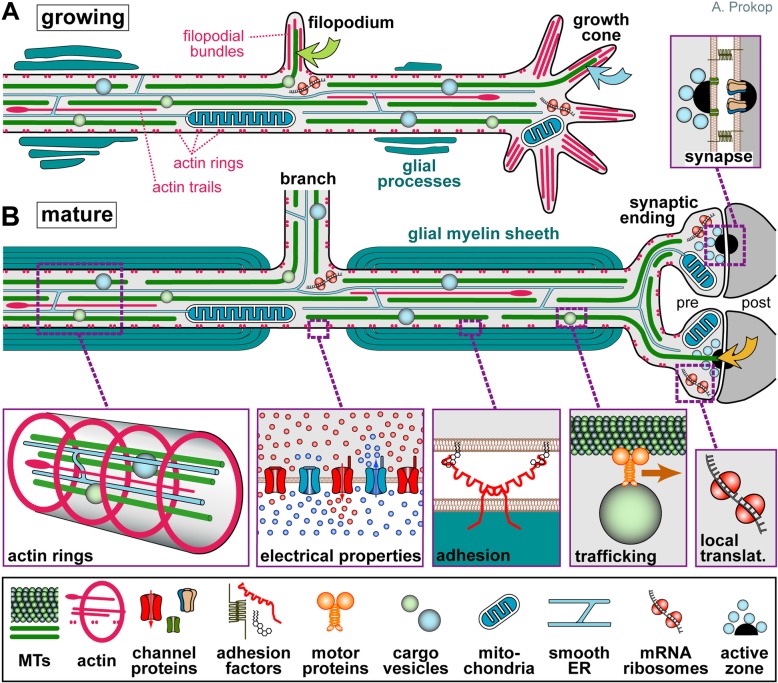
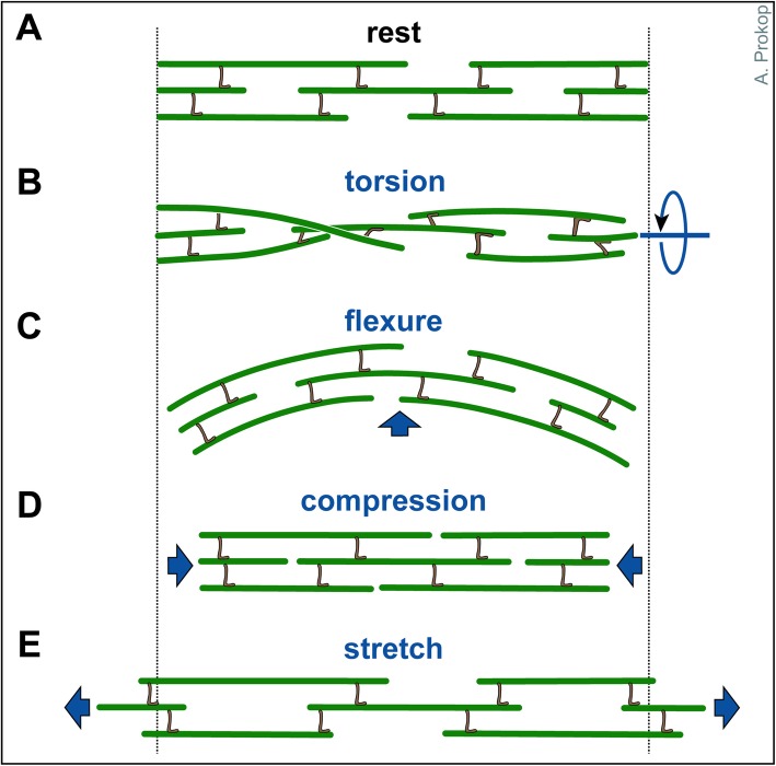
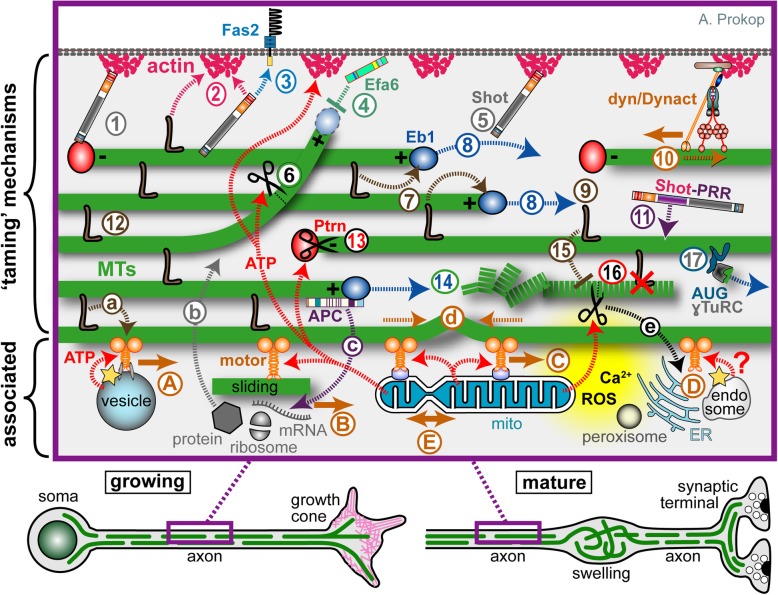
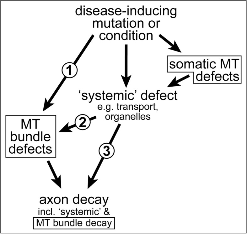
Axons are the slender, cable-like, up to meter-long projections of neurons that electrically wire our brains and bodies. In spite of their challenging morphology, they usually need to be maintained for an organism's lifetime. This makes them key lesion sites in pathological processes of ageing, injury and neurodegeneration. The morphology and physiology of axons crucially depends on the parallel bundles of microtubules (MTs), running all along to serve as their structural backbones and highways for life-sustaining cargo transport and organelle dynamics. Understanding how these bundles are formed and then maintained will provide important explanations for axon biology and pathology. Currently, much is known about MTs and the proteins that bind and regulate them, but very little about how these factors functionally integrate to regulate axon biology. As an attempt to bridge between molecular mechanisms and their cellular relevance, we explain here the model of local axon homeostasis, based on our own experiments in Drosophila and published data primarily from vertebrates/mammals as well as C. elegans. The model proposes that (1) the physical forces imposed by motor protein-driven transport and dynamics in the confined axonal space, are a life-sustaining necessity, but pose a strong bias for MT bundles to become disorganised. (2) To counterbalance this risk, MT-binding and -regulating proteins of different classes work together to maintain and protect MT bundles as necessary transport highways. Loss of balance between these two fundamental processes can explain the development of axonopathies, in particular those linking to MT-regulating proteins, motors and transport defects. With this perspective in mind, we hope that more researchers incorporate MTs into their work, thus enhancing our chances of deciphering the complex regulatory networks that underpin axon biology and pathology.

Keywords: Drosophila; actin; axons; cytoskeleton; microtubules; neurodegeneration.


Conflict of interest statement

The authors declare that they have no competing interests.

Figures

Fig. 1
Fig. 1
Specific properties of axons. Axons during the growth cone stage are shown in (a) and after synaptic maturation in (b), differing primarily in certain stage-specific specialisations including growth cones, synapses, electrical properties and glial interactions (here myelination [389, 392]). The core machinery in the axon shaft can be expected to be similar at both stages: parallel continuous bundles of extended but discontinuous MTs run all along axons serving as a structural backbone (see Fig. 2), a transport highway for axonal trafficking (driven by motor proteins), and a source for 'off-track' MTs contributing to morphogenetic processes including branch formation, directed axon growth and synapse formation/plasticity (green, orange, blue curved arrows); MT bundles are interspersed with longitudinal actin trails [18, 24], continuous networks of (smooth) endoplasmic reticulum [44, 393], and other membranous organelles including mitochondria [45]; axonal membranes display regularly spaced periodic rings of cortical actin [20, 21], a high number of ion-specific channel proteins and transporters to conduct nerve impulses [394], as well as adhesions with external structures including fasciculating parallel axons (not shown), glial processes [395] and synaptic partner cells [396]; a degree of independence from cell-body derived proteins is provided by local translation machinery [–399] or supply from surrounding glia cells (not shown; [–403]). Note that the axon diameter in the region between glia cells in B (referred to as Node of Ranvier) usually has a much smaller diameter than the rest of the axon [1]
Fig. 2
Fig. 2
Axonal response to mechanical challenges. Continuous bundles of discontinuous MTs which are flexibly cross-linked (likely involving slip-bonds) are thought to provide a structural element that can respond to different forms of mechanical impact (as indicated in blue)
Fig. 3
Fig. 3
An interactome of MT-regulating and -associated mechanisms expected to contribute within the model of local axon homeostasis. Developing and mature neurons are shown at the bottom indicating that the close-up (magenta frame) might apply in both contexts. 1-16) Potential mechanisms that can 'tame' MTs into bundled conformation: MT polymerisation (blue stippled arrows) is driven by molecular machinery centred on Eb1 (blue balls), further influenced by the tubulin supply machinery (not shown) and shaft-binding proteins (7); polymerisation generates new MTs required for bundle formation (8) and turn-over (14); to integrate into bundles, extending MTs require guidance via actin-Eb1 cross-linkage along the axonal surface (5; Shot) or along pre-existing MTs through MT-MT cross-linkers (9; brown L). The same or other cross-linkers provide the structural glue that holds MT bundles together (12; brown L); some of them can also bind to actin (2), they protect from (or recruit) MT severing activity (15), and influence motor protein dynamics (a). MTs which have escaped any cross-linkage are inhibited by cortical collapse factors when approaching the axonal surface (4; Efa6) or by MT-severing factors at MT-MT cross-points (6). The bundled MTs are discontinuous; their free minus ends are stabilised by CAMSAP/Patronin (Ptrn) together with katanin (black scissors; 13), whereas non-polymerising MT plus ends are stabilised by other factors (not shown; e.g. CLASP or the Dynactin subunit p150/Glued [404, 405]). The dynein/Dynactin complex is believed to link cortical actin to MT bundles and drive them anterogradely (10), whereas Ptrn at minus ends may anchor MTs via spectraplakins to the axon cortex (1); spectraplakins may also link MTs directly to cortical actin (2) or to transmembrane receptors (3), and they are expected to perform further, still unexplored actin-independent bundle-promoting roles through their PRR domains (11). Tear-and-wear damages MTs (dashed green line), potentially affecting interaction with MT-binding proteins (16; red X); MT severing proteins might selectively eliminate such MTs (16; scissors), or MTs undergo repair (not shown). Nucleation of MTs (17) is mediated by ɣTuRC directionally anchored to MT lattices via the augmin/HAUS complex (AUG). A-E) Mechanisms closely 'associated' with MT bundles: MT-associated motor proteins ('motor', solid orange arrows) drive axonal transport of (protein-loaded) vesicles (A), cytoplasmic factors including proteins, translational machinery (ribosomes) or RNAs (B), move other MTs (B, sliding), and position/rearrange organelles including mitochondria (C, mitos), endoplasmic reticulum, peroxisomes and endosome (D) - and this likely includes mitochondrial fission and fusion (E). a-e) The motor-associated functions all act downstream of MT bundles because they require them to walk on; but they also act upstream: for example, the forces they generate (stippled orange arrows) are the potential cause for MT disorganisation (buckling shown in d); transport delivers important regulators and building blocks for bundle-maintaining processes (b); the proper regulation of organelles/endocytic compartments provides systemic factors that can orchestrate MT bundle-taming mechanisms, including intracellular free calcium or reactive oxygen species (Ca2+, ROS; yellow cloud [202, 203]) as well as ATP required for many processes including actin dynamics, MT severing and MT motor activity (red stippled arrows; note that vesicular transport uses glycolysis to generate its own ATP; yellow star); vice versa, the MT severer spastin also regulates the ER through ATP-independent mechanisms (e), and MT-associated proteins (APC) regulate local translation events (c)
Fig. 4
Fig. 4
MT bundle defects as cause or consequence of axon decay. 1) Disease-inducing mutations/conditions can affect a MT bundle regulator (e.g. dystonin [90]), thus causing MT bundle defects first which, in turn, can trigger axon decay. 2) Disease-inducing mutations/conditions can affect systemic factors which, in turn cause MT bundle defects as an intermediate causative step in the cascade leading to axon decay (e.g. axonal transport fails, leading to MT bundle defects which then contribute to axon decay, as is the case in Alzheimer's disease or ALS [302, 406, 407]); this may occur even if MT regulators are affected, but these regulators mainly act in the cell body (e.g. dysregulation of the Golgi [408]). 3) MT bundle deterioration may be a mere consequence of axon decay, although this case will be difficult to disentangle from option 2, since MT bundle disintegration and axonal disassembly may occur in parallel, as observed in developmental or injury-induced axon degeneration [–411]). All MT-related phenotypes in this graph are indicated with a frame
Fig. 5
Fig. 5
Disorganisation of axonal MTs upon loss of different MT regulators in Drosophila primary neurons. a Normal neuron (wild-type, wt) with soma (asterisk), axon shaft (curved arrow) and growth cone (tip of most distal MT indicated by arrow head). b Eb15 mutant neuron where the area of MT disorganisation is framed by a red stippled box and shown as close-up on the right. c-e Similar close-ups shown for Efa6GX6[w-], Khc27 and shot3 mutant neurons. Note that the four mutated factors perform fundamentally different molecular functions, with Eb1 being a MT plus-end binder ('8' in Fig. 3), Efa6 a cortical collapse factor ('4' in Fig. 3), Khc a kinesin-1 motor protein ('A-E' in Fig. 3) and Shot a multi-functional cross-linker ('1-3, 5, 11' in Fig. 3). All neurons were derived from wild-type or homozygous mutant embryos, mechanically and chemically dissociated, kept for 7 days in pre-culture in a centrifuge tube to deplete any maternal gene product, mechanically and chemically dissociated again, cultured on concanavalin A-coated glass coverslips for 1day at 21°C, fixed and stained with anti-α-tubulin (DM1A, Sigma; procedures detailed elsewhere: [78]); images were taken by A.V. using STED (stimulated emission depletion) microscopy. Scale bar in A represents 10 μm for the two neurons and 4 μm in close-ups
Fig. 6
Fig. 6
A molecular perspective of microtubule properties. a Cross-section of a MT with 14 protofilaments (PF) and lateral view of a 13 PF MT, both in B-lattice configuration, where α-tubulins make lateral bonds with α-tubulins and ß with ß, except at the seam (magenta line: seam; dashed red line: PF). b Close-up of an α/ß-tubulin heterodimer showing the various post-translational modification sites as indicated; note that the GTP of ß-tubulin in lattices is usually hydrolysed (GDP). c A 13 PF MT (top), cut open at the seam and rolled out (bottom); the yellow line shows the diameter, the white line follows the helical rise of laterally bonded tubulins; in 13 PF MTs, tubulins are precisely aligned at the seam (yellow arrow head) but shifted by three positions (3-start helix). d When deviating from the 13 PF prototype, tubulins are misaligned at the seam (orange arrow head); when forced into alignment, the PFs skew (deviation of the magenta line from the white stippled line), causing a super-twist of the MT as described by the 'lattice accommodation model' [98, 412]; for certain PF numbers, MTs can form two alternative alignments, of which usually the version with the lower helix start value (left) has a left-handed super-twist, whereas the higher value is right-handed [98]. e MTs behave like rigid rods with a persistence length of up to 10 mm, but can be bent down to diameters of curvature of ~1μm before they break; it has been reported that their cross-sectional profile may flatten above a certain threshold (black arrow head), thus softening the tube. f Lattices of GDP-tubulin are 1-3% shorter than MTs that were polymerised with the non-hydrolysable GTP analogue GMPCPP, or stabilised with taxol (orange structure binding α-tubulin in a 1:1 ratio, according to [413]); binding of kinesin-1 causes similar lengthening of tubulin (and additional compactions in the tubulin structure: yellow stars) which may cause cooperative binding of further kinesins and induce curvature if occurring only on one side of the MT; in extended taxol-bound MTs, bending forces were suggested to change tubulins on the concave side into their short conformation as an energetically favoured condition. For further references see main text

Similar articles

Cited by

References

    1. Hoffman PN. Review : The Synthesis, Axonal Transport, and Phosphorylation of Neurofilaments Determine Axonal Caliber in Myelinated Nerve Fibers. Neuroscientist. 1995;1:76–83. doi: 10.1177/107385849500100204. - DOI
    1. Bichenback J. International perspectives on spinal cord injury. WHO, ISCOS: Switzerland; 2013.
    1. Tedeschi A, Bradke F. Spatial and temporal arrangement of neuronal intrinsic and extrinsic mechanisms controlling axon regeneration. Curr Opin Neurobiol. 2016;42:118–127. doi: 10.1016/j.conb.2016.12.005. - DOI - PubMed
    1. Medana IM, Esiri MM. Axonal damage: a key predictor of outcome in human CNS diseases. Brain. 2003;126:515–530. doi: 10.1093/brain/awg061. - DOI - PubMed
    1. Gaetz M. The neurophysiology of brain injury. Clin Neurophysiol. 2004;115:4–18. doi: 10.1016/S1388-2457(03)00258-X. - DOI - PubMed

Publication types

LinkOut - more resources