. 2019 Nov 9;7(1):175.
doi: 10.1186/s40478-019-0833-0.

The level of activity of the alternative lengthening of telomeres correlates with patient age in IDH-mutant ATRX-loss-of-expression anaplastic astrocytomas

Collaborators, Affiliations

The level of activity of the alternative lengthening of telomeres correlates with patient age in IDH-mutant ATRX-loss-of-expression anaplastic astrocytomas

Nathalie Grandin et al. Acta Neuropathol Commun. .

Abstract

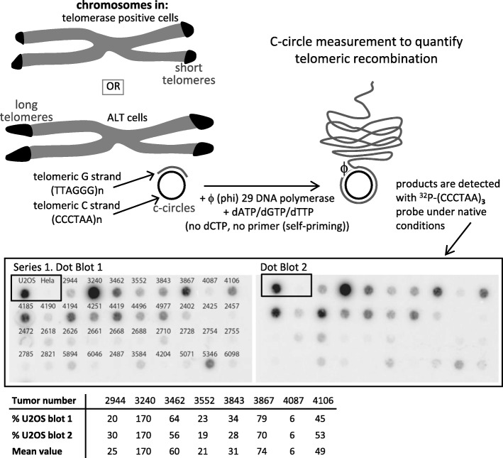
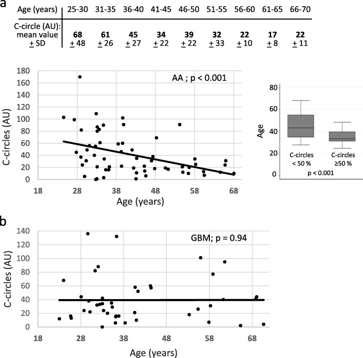
All cancer cells need to maintain functional telomeres to sustain continuous cell division and proliferation. In human diffuse gliomas, functional telomeres are maintained due either to reactivation of telomerase expression, the main pathway in most cancer types, or to activation of a mechanism called the alternative lengthening of telomeres (ALT). The presence of IDH1/2 mutations (IDH-mutant) together with loss of ATRX expression (ATRX-lost) are frequently associated with ALT in diffuse gliomas. However, detection of ALT, and a fortiori its quantification, are rarely, if ever, measured in neuropathology laboratories. We measured the level of ALT activity using the previously described quantitative "C-circle" assay and analyzed it in a well characterized cohort of 104 IDH-mutant and ATRX-lost adult diffuse gliomas. We report that in IDH-mutant ATRX-lost anaplastic astrocytomas, the intensity of ALT was inversely correlated with age (p < 0.001), the younger the patient, the higher the intensity of ALT. Strikingly, glioblastomas having progressed from anaplastic astrocytomas did not exhibit this correlation. ALT activity level in the tumor did not depend on telomere length in healthy tissue cells from the same patient. In summary, we have uncovered the existence, in anaplastic astrocytomas but not in glioblastomas with the same IDH and ATRX mutations, of a correlation between patient age and the level of activity of ALT, a telomerase-independent pathway of telomere maintenance.

Keywords: ATRX loss of expression; Alternative lengthening of telomeres; Anaplastic astrocytoma; IDH1/2 mutations; Secondary glioblastoma.


Conflict of interest statement

The authors declare that they have no competing interests.

Figures

Fig. 1
Fig. 1
Measurement of the level of ALT activity levels in human diffuse gliomas. Top panels illustrate the principle of the ALT C-circle assay [17] and describe its general steps. ALT cells have very long telomeres that have been amplified mainly by homologous recombination that generates partially single-stranded extra-chromosomal circles. Genomic DNA prepared from tumor samples is then incubated with the Phi29 DNA polymerase that specifically amplifies this telomeric DNA. Middle panel illustrates ALT-specific signals measured in tumor DNA samples using this assay, which were detected here on dot blots hybridized with a telomeric 32P-labeled probe. Genomic DNAs from HeLa (telomerase positive) and U2OS (ALT positive) cells were also probed, representing negative and posititve controls for the C-circle assay, respectively. These assays were systematically performed in duplicates and here dot blot 2, on the right, was loaded with the same tumor samples as dot blot 1, on the left, to insure for reproducibility. Bottom table illustrates examples of duplicate numbers obtained for each of the indicated tumors, real signals of which are represented in the middle panel above. The C-circle score was determined after calculating the intensity of the signal relative to that of the ALT positive U2OS cell line, designated to be 100 arbitrary units (AU). Note that the C-circle assays were performed on representative samples, including those from the two patient groups analyzed in the present study
Fig. 2
Fig. 2
Correlation between the level of ALT activity, expressed as the percentage of radioactive signal with respect to the ALT signal recorded in genomic DNA from U2OS cells, designated to be 100 arbitrary units (AU), and patient age in anaplastic astrocytomas, AA (a, top and bottom left) and secondary glioblastomas multiform, GBM (b). Patient age was considered at the time of first surgery. a bottom right: According to statistical distribution, we arbitrarily defined two categories of anaplastic astrocytoma (AA) patients with C-circle score < or > 50 AU. This graphical representation also clearly shows the ALT intensity/patient age correlation
Fig. 3
Fig. 3
Detection of ultra-bright telomeric foci by Telo-FISH in paraffin-embedded sections of tumors, together with detection of DNA by DAPI in the same section, as indicated (40X magnification). The upper chart indicates the mean number of telomeric foci per nucleus, as well as the percentage of nuclei with ultra-bright foci of an intensity over 40 pixels (px), the rate of C-circles and the diagnostic. C-circles values are expressed in arbitrary units (AU; see Materials and methods). The images are representative pictures of tumors; OD: oligodendroglioma; GBM IDHmt: IDH-mutant secondary glioblastoma; AA: anaplastic astrocytoma. Note that the photograph representing the OD tumor (picture a) was taken at a much higher exposure, as indicated, to show that the Telo-FISH signals are indeed present, but are much fainter than in the GBM and AA tumors. For both the OD tumor (a) and AA tumor (b) shown in the upper panels, lower magnifications are provided in the lower panels, as indicated (larger field)

References

    1. Barthel FP, Wei W, Tang M, Martinez-Ledesma E, Hu X, Amin SB, Akdemir KC, Seth S, Song X, Wang Q, et al. Systematic analysis of telomere length and somatic alterations in 31 cancer types. Nat Genet. 2017;49:349–357. doi: 10.1038/ng.3781. - DOI - PMC - PubMed
    1. Broccoli D, Godley LA, Donehower LA, Varmus HE, de Lange T. Telomerase activation in mouse mammary tumors: lack of detectable telomere shortening and evidence for regulation of telomerase RNA with cell proliferation. Mol Cell Biol. 1996;16:3765–3772. doi: 10.1128/mcb.16.7.3765. - DOI - PMC - PubMed
    1. Bryan TM, Englezou A, Dalla-Pozza L, Dunham MA, Reddel RR. Evidence for an alternative mechanism for maintaining telomere length in human tumors and tumor-derived cell lines. Nat Med. 1997;3:1271–1274. doi: 10.1038/nm1197-1271. - DOI - PubMed
    1. Cancer Genome Atlas Research N. Brat DJ, Verhaak RG, Aldape KD, Yung WK, Salama SR, Cooper LA, Rheinbay E, Miller CR, Vitucci M, et al. Comprehensive, integrative genomic analysis of diffuse lower-grade Gliomas. N Engl J Med. 2015;372:2481–2498. doi: 10.1056/NEJMoa1402121. - DOI - PMC - PubMed
    1. Cerone MA, Autexier C, Londono-Vallejo JA, Bacchetti S. A human cell line that maintains telomeres in the absence of telomerase and of key markers of ALT. Oncogene. 2005;24:7893–7901. doi: 10.1038/sj.onc.1208934. - DOI - PubMed

Publication types