. 2019 Dec 10;13(6):960-969.
doi: 10.1016/j.stemcr.2019.10.006. Epub 2019 Nov 7.

Effects of Spaceflight on Human Induced Pluripotent Stem Cell-Derived Cardiomyocyte Structure and Function

Affiliations

Effects of Spaceflight on Human Induced Pluripotent Stem Cell-Derived Cardiomyocyte Structure and Function

Alexa Wnorowski et al. Stem Cell Reports. .

Abstract

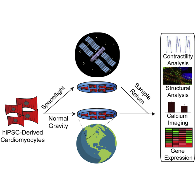
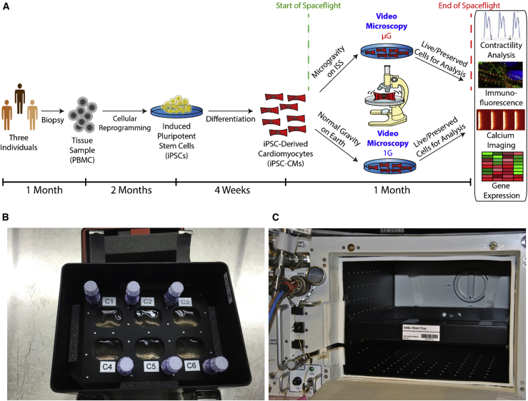
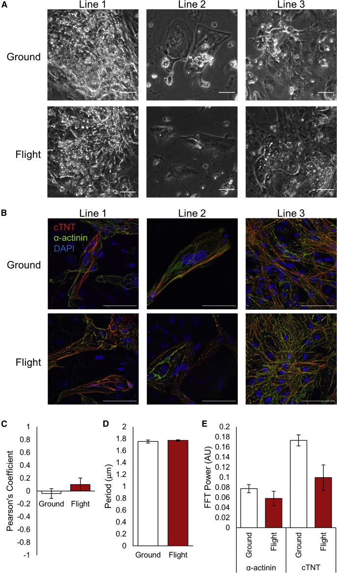
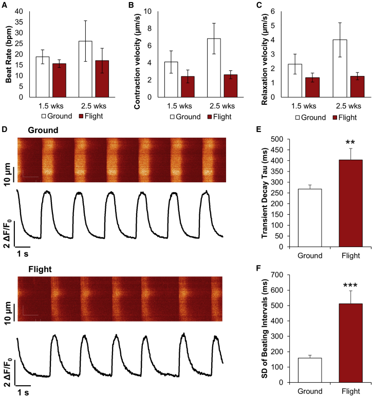
With extended stays aboard the International Space Station (ISS) becoming commonplace, there is a need to better understand the effects of microgravity on cardiac function. We utilized human induced pluripotent stem cell-derived cardiomyocytes (hiPSC-CMs) to study the effects of microgravity on cell-level cardiac function and gene expression. The hiPSC-CMs were cultured aboard the ISS for 5.5 weeks and their gene expression, structure, and functions were compared with ground control hiPSC-CMs. Exposure to microgravity on the ISS caused alterations in hiPSC-CM calcium handling. RNA-sequencing analysis demonstrated that 2,635 genes were differentially expressed among flight, post-flight, and ground control samples, including genes involved in mitochondrial metabolism. This study represents the first use of hiPSC technology to model the effects of spaceflight on human cardiomyocyte structure and function.

Keywords: calcium imaging; cardiology; cardiomyocytes; heart; induced pluripotent stem cells; metabolism; microgravity; modeling; spaceflight; stem cell.


Figures

None
Graphical abstract
Figure 1
Figure 1
Evaluating the Effects of Spaceflight on hiPSC-CM Structure and Function (A) Timeline for experiment. Tissue samples were collected from three individuals and used to generate human induced pluripotent stem cell-derived cardiomyocytes (hiPSC-CMs). The hiPSC-CMs were plated in fully enclosed 6-well plates optimized for microgravity (“BioCells”) and sent to the International Space Station (ISS) for culture and live imaging for ∼1 month. Ground controls with the same hiPSC-CM lines and hardware were maintained at Stanford University. Media exchanges and imaging were strictly scheduled so that the only significant difference in cell environment was spaceflight. After sample return from the ISS, cellular phenotypes were evaluated using gene expression, immunofluorescence, calcium imaging, and contractility analyses. (B) A BioCell in its environmental control habitat (“PHAB”). (C) The interior of the Space Automated Bioproduct Laboratory (SABL), the incubator used to maintain the cells on the ISS.
Figure 2
Figure 2
hiPSC-CMs Demonstrate No Overt Changes in Cell Morphology or Sarcomere Structure after Return from Spaceflight (A) Representative phase-contrast images of live-return hiPSC-CMs from each cell line 3 days after sample return from the ISS, prior to paraformaldehyde fixation. (B) Immunofluorescence of flight and ground control samples from each cell line showing sarcomeric proteins cardiac troponin T (cTnT) and α-actinin, and nuclear stain DAPI. Scale bars represent 50 μm. (C–E) Pearson's coefficient (C), period (D), and fast Fourier transform (E) power of cTnT and α-actinin signals along sarcomere lines for ground and flight samples (arbitrary units). N = 3 lines, n = 2–3 images per line, with 8–15 sarcomeres analyzed per image. Error bars represent standard error of the mean (SEM).
Figure 3
Figure 3
hiPSC-CM Contraction and Calcium Handling Are Altered by Spaceflight (A–C) Beat rate in beats per minute (bpm) (A), contraction velocity (B), and relaxation velocity (C) for ground control and flight hiPSC-CMs after 1.5 and 2.5 weeks of culture on the ISS. N = 3 lines, n = 1–2 biological replicates per line, with 1–4 videos per sample. (D) Representative calcium transients for ground and flight conditions, measured 3 days after live return from the ISS. (E and F) Transient decay tau (E) and standard deviation of beating intervals (F) from calcium transients. N = 103 and 34 cells in ground and flight groups, respectively. ∗∗p < 0.01 and ∗∗∗p < 0.001 versus ground control. Error bars represent SEM. See also Figure S1; Videos S1 and S2.
Figure 4
Figure 4
hiPSC-CM RNA Expression Profiles Are Altered by Spaceflight RNA-sequencing data comparing flight samples preserved in RNAlater after ∼4.5 weeks in microgravity, post-flight samples preserved 10 days post return after ∼5.5 weeks in microgravity, and ground control samples preserved at the same time as post-flight samples. (A) Heatmap displaying 2,635 genes differentially expressed among the three groups with p ≤ 0.05. (B–D) Expression of genes related to (B) calcium handling and contraction, (C) hypertrophy, and (D) metabolism, with p ≤ 0.05. (E) Group enrichment scores for top functional annotation clusters (enrichment score ≥2 for at least one group) determined using the Database for Annotation, Visualization, and Integrated Discovery (DAVID) for genes differentially expressed between the indicated groups with p ≤ 0.05 based on a two-tailed Student’s t test. (F) Venn diagram demonstrating differentially expressed genes for each comparison with p ≤ 0.05 based on a two-tailed Student’s t test. See also Figure S2 and Tables S1, S2, and S3.

References

    1. Agrawal R., Agrawal N., Koyani C.N., Singh R. Molecular targets and regulators of cardiac hypertrophy. Pharmacol. Res. 2010;61:269–280. - PubMed
    1. Azakie A., Fineman J.R., He Y. Sp3 inhibits Sp1-mediated activation of the cardiac troponin T promoter and is downregulated during pathological cardiac hypertrophy in vivo. Am. J. Physiol. Circ. Physiol. 2006;291:H600–H611. - PubMed
    1. Baio J., Martinez A.F., Silva I., Hoehn C.V., Countryman S., Bailey L., Hasaniya N., Pecaut M.J., Kearns-Jonker M. Cardiovascular progenitor cells cultured aboard the International Space Station exhibit altered developmental and functional properties. NPJ Microgravity. 2018;4:13. - PMC - PubMed
    1. Becker J.L., Souza G.R. Using space-based investigations to inform cancer research on Earth. Nat. Rev. Cancer. 2013;13:315–327. - PubMed
    1. Bergmann O., Bhardwaj R.D., Bernard S., Zdunek S., Barnabé-Heider F., Walsh S., Zupicich J., Alkass K., Buchholz B.A., Druid H. Evidence for cardiomyocyte renewal in humans. Science. 2009;324:98–102. - PMC - PubMed

Publication types