Review
. 2019 Nov-Dec;10(6):1082-1093.
doi: 10.1016/j.jcot.2019.07.004. Epub 2019 Jul 13.

Vitamin D for skeletal and non-skeletal health: What we should know

Affiliations
Review

Vitamin D for skeletal and non-skeletal health: What we should know

Nipith Charoenngam et al. J Clin Orthop Trauma. 2019 Nov-Dec.

Abstract

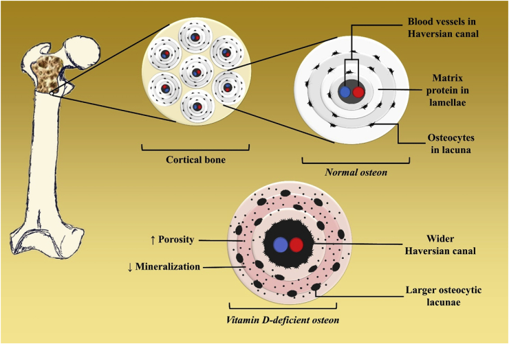
Vitamin D plays an essential role in regulating calcium and phosphate metabolism and maintaining a healthy mineralized skeleton. Humans obtain vitamin D from sunlight exposure, dietary foods and supplements. There are two forms of vitamin D: vitamin D3 and vitamin D2. Vitamin D3 is synthesized endogenously in the skin and found naturally in oily fish and cod liver oil. Vitamin D2 is synthesized from ergosterol and found in yeast and mushrooms. Once vitamin D enters the circulation it is converted by 25-hydroxylase in the liver to 25-hydroxyvitamin D [25(OH)D], which is further converted by the 25-hydroxyvitamin D-1α-hydroxylase in the kidneys to the active form, 1,25-dihydroxyvitamin D [1,25(OH)2D]. 1,25(OH)2D binds to its nuclear vitamin D receptor to exert its physiologic functions. These functions include: promotion of intestinal calcium and phosphate absorption, renal tubular calcium reabsorption, and calcium mobilization from bone. The Endocrine Society's Clinical Practice Guideline defines vitamin D deficiency, insufficiency, and sufficiency as serum concentrations of 25(OH)D of <20 ng/mL, 21-29 ng/mL, and 30-100 ng/mL, respectively. Vitamin D deficiency is a major global public health problem in all age groups. It is estimated that 1 billion people worldwide have vitamin D deficiency or insufficiency. This pandemic of vitamin D deficiency and insufficiency is attributed to a modern lifestyle and environmental factors that restrict sunlight exposure, which is essential for endogenous synthesis of vitamin D in the skin. Vitamin D deficiency is the most common cause of rickets and osteomalacia, and can exacerbate osteoporosis. It is also associated with chronic musculoskeletal pain, muscle weakness, and an increased risk of falling. In addition, several observational studies observed the association between robust levels of serum 25(OH)D in the range of 40-60 ng/mL with decreased mortality and risk of development of several types of chronic diseases. Therefore, vitamin D-deficient patients should be treated with vitamin D2 or vitamin D3 supplementation to achieve an optimal level of serum 25(OH)D. Screening of vitamin D deficiency by measuring serum 25(OH)D is recommended in individuals at risk such as patients with diseases affecting vitamin D metabolism and absorption, osteoporosis, and older adults with a history of falls or nontraumatic fracture. It is important to know if a laboratory assay measures total 25(OH)D or only 25(OH)D3. Using assays that measure only 25(OH)D3 could underestimate total levels of 25(OH)D and may mislead physicians who treat patients with vitamin D2 supplementation.

Keywords: Vitamin D.


Figures

Fig. 1
Fig. 1
UV radiation therapy for rickets. (A) Photograph from the 1920s of a child with rickets being exposed to artificial UV radiation. (B) Radiographs demonstrating florid rickets of the hand and wrist (left). The same hand and wrist taken after a course of treatment with 1-h UV radiation 2 times/week for 8 week showing mineralization of the carpal bones and epiphyseal plates (right). Holick, copyright 2006. Reproduced with permission.
Fig. 2
Fig. 2
Schematic representation of the synthesis and metabolism of vitamin D for skeletal and nonskeletal function. 1-OHase = 25-hydroxyvitamin D-1α-hydroxylase; 24-OHase = 25-hydroxyvitamin D-24-hydroxylase; 25(OH)D = 25-hydroxyvitamin D; 1,25(OH)2D = 1,25-dihydroxyvitamin D; CaBP = calcium-binding protein; CYP27B1, Cytochrome P450–27B1; DBP = vitamin D–binding protein; ECaC = epithelial calcium channel; FGF-23 = fibroblast growth factor-23; PTH = parathyroid hormone; RANK = receptor activator of the NF-kB; RANKL = receptor activator of the NF-kB ligand; RXR = retinoic acid receptor; TLR2/1 = Toll-like receptor 2/1; VDR = vitamin D receptor; vitamin D = vitamin D2 or vitamin D3. Copyright Holick 2013, reproduced with permission.
Fig. 3
Fig. 3
In vitamin D-deficient bone, increased parathyroid hormone induces the osteoblast to express receptor activator of NF-kB ligand (RANKL) on their cell surface and to secrete soluble RANKL into the extracellular matrix. Both surface RANKL and soluble RANKL interact with receptor activator of NF-kB on the surface of osteoclast precursor cells which are differentiated from macrophage-colony forming unit (M-CFU). RANK-RANKL interaction leads to the differentiation of osteoclast precursor cells into multinucleated osteoclasts which are then activated to exert its bone resorbing activity.
Fig. 4
Fig. 4
Activation of receptor activator NF-kB (RANK) by receptor activator NF-kB ligand (RANKL) leads to osteoclast differentiation and osteoclast activation. It promotes bone resorbing activity of the osteoclast in the lacuna by inducing its secretion of acids including citric acid, lactic acid, hydrochloric acid, and enzymes including cathepsins and matrix metalloproteinase (MMP), leading to a release in collagen fragment, calcium and phosphate from the bone into the extracellular matrix.
Fig. 5
Fig. 5
When serum 25-hydroxyvitamin D (25(OH)D) is less than 30 ng/mL, there is a significant decrease in intestinal calcium and phosphate absorption. This causes a decrease in serum ionized calcium concentration and subsequent secondary hyperparathyroidism. Elevated parathyroid hormone (PTH) induces differentiation of preosteoclast into mature osteoclast leading to increased osteoclast activity. This results in increased bone resorption, loss of bone mineral and matrix, and subsequent low bone mass and osteoporosis. In addition, PTH displays a phosphaturic effect leading to an increase in urinary phosphate excretion. Urinary phosphate loss and decreased intestinal phosphate absorption due to vitamin D deficiency [25(OH)D < 20 ng/mL] contribute to inadequate calcium-phosphate product, thereby resulting in defective bone mineralization and development of rickets and osteomalacia.
Fig. 6
Fig. 6
Normal bone histology with normal trabeculae and normal bone mass is demonstrated in the left picture. Histology of bone with low bone mass/osteoporosis demonstrating thin trabeculae, poor connectivity and low bone mass is shown in the middle picture. Histology of bone with rickets/osteomalacia demonstrating accumulated osteoid (red areas around black mineralized trabeculae) is shown in the right picture.
Fig. 7
Fig. 7
Bone consists of two major components, including cortical bone which is a major determinant of bone strength and resistance to fracture, and trabecular bone which acts as bridges built inside the cortical bone that intensifies bone strength. Cortical bone is a built of cylinders called osteons. Each osteon is composed of bone collagen matrix protein arranged concentrically in lamellae around a Haversian canal containing venous (blue) and arterial (red) blood vessels. Osteocytes and osteoclasts are located in lacunae between each lamellae. Vitamin D-deficient osteons displayed larger lacunae, wider Haversian canal due to the PTH induced increase in numbers and activity of osteoclasts thereby increasing the porosity. In addition, there is defect in osteoid mineralization (light pink area) compared with those of normal bone.
Fig. 8
Fig. 8
Skeletal deformities observed in rickets. (A) Photograph from the 1930s of a sister (left) and brother (right), aged 10 months and 2.5 years, respectively, showing enlargement of the ends of the bones at the wrist, carpopedal spasm, and a typical “Taylorwise” posture of rickets. (B) The same brother and sister 4 years later, with classic knock-knees and bow legs, growth retardation, and other skeletal deformities. Holick, copyright 2006. Reproduced with permission.

References

    1. Hess A.F. Henry Kimpton; London W.C.: 1930. Rickets Including Osteomalacia and Tetany. 263, High Holborn; p. 485. xv +
    1. Still G.F. London EOUP, Humphrey Milford; 1931. The History of Pediatrics. The Progress of the Study of Disease of Children up to the End of XVIIIth Century.
    1. Holick M.F. Resurrection of vitamin D deficiency and rickets. J Clin Invest. 2006;116(8):2062–2072. - PMC - PubMed
    1. Huldschinsky K. Heilung von Rachitis durch künstliche Höhensonne. Dtsch med Wochenschr. 1919;45(26):712–713.
    1. Rajakumar K., Greenspan S.L., Thomas S.B., Holick M.F. SOLAR ultraviolet radiation and vitamin D: a historical perspective. Am J Public Health. 2007;97(10):1746–1754. - PMC - PubMed