Review
. 2019 Oct 25:10:1264.
doi: 10.3389/fphar.2019.01264. eCollection 2019.

Recent Trends of the Bio-Inspired Nanoparticles in Cancer Theranostics

Affiliations
Review

Recent Trends of the Bio-Inspired Nanoparticles in Cancer Theranostics

Vijay Sagar Madamsetty et al. Front Pharmacol. .

Abstract

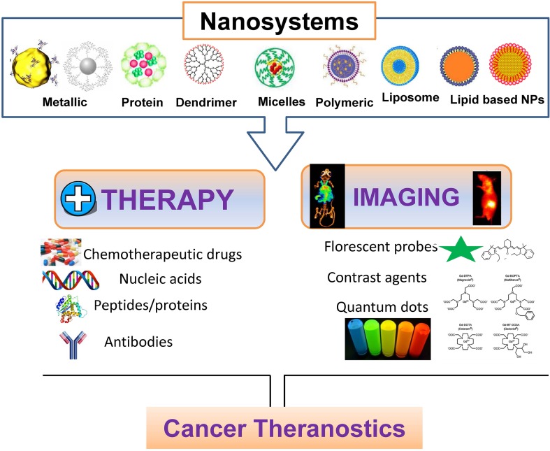
In recent years, various nanomaterials have emerged as an exciting tool in cancer theranostic applications due to their multifunctional property and intrinsic molecular property aiding effective diagnosis, imaging, and successful therapy. However, chemically synthesized nanoparticles have several issues related to the cost, toxicity and effectiveness. In this context, bio-inspired nanoparticles (NPs) held edges over conventionally synthesized nanoparticles due to their low cost, easy synthesis and low toxicity. In this present review article, a detailed overview of the cancer theranostics applications of various bio-inspired has been provided. This includes the recent examples of liposomes, lipid nanoparticles, protein nanoparticles, inorganic nanoparticles, and viral nanoparticles. Finally, challenges and the future scopes of these NPs in cancer therapy and diagnostics applications are highlighted.

Keywords: bio-inspired nanoparticles; cancer; clinical trials; imaging; nanomedicine; theranostics.


Figures

Figure 1
Figure 1
Nanotheranostics: polymeric, lipid-based and metallic nanomaterials for cancer theranostics.
Figure 2
Figure 2
Schematic illustration of various HAS based nanoparticles. (A) HAS-Gd-IR825 (Chen et al., 2014), (B) P@-Gem-HSA (Yu et al., 2017), and (C) HSA/dc-IR825/GA complex (Gao et al., 2019).
Figure 3
Figure 3
Over all presentation for synthesis, characterization and biomedical applications (diagnostic, anticancer antibacterial applications) of biosynthesized silver nanoparticles (b-AgNPs) using Olax Scandens leaf extract. Reprinted with permission from (Mukherjee et al., 2014).
Figure 4
Figure 4
Live/dead staining of A431 cells after 800 nm laser irradiation for 10 min at power = 36 mW. Nanoparticle concentration in panels (AC) and (D) are 200, 100, 50, and 0 μg/ml respectively. Decreasing amounts of red fluorescence is obtained with lower concentration of nanoparticles which indicates the photothermal activity of the nanoparticles in cell killing. (E) Live/dead staining of A431 cells treated with 100 μg/ml nanoparticle concentration without laser treatment. Reprinted with permission from (Fazal et al., 2014). Copyright @ American Chemical Society.

References

    1. Al-Jamal W. T., Al-Jamal K. T., Tian B., Cakebread A., Halket J. M., Kostarelos K. (2009). Tumor Targeting of Functionalized Quantum Dot–Liposome Hybrids by Intravenous Administration. Mol. Pharmaceut. 6, 520–530. 10.1021/mp800187d - DOI - PubMed
    1. An F.-F., Zhang X.-H. (2017). Strategies for Preparing Albumin-based Nanoparticles for Multifunctional Bioimaging and Drug Delivery. Theranostics 7, 3667–3689. 10.7150/thno.19365 - DOI - PMC - PubMed
    1. Ana L., Nuno G. A., Andreia C. G., Artur C.-P. (2016). Albumin-Based Nanodevices as Drug Carriers. Curr. Pharm. Des. 22, 1371–1390. 10.2174/1381612822666160125114900 - DOI - PubMed
    1. Au - Wen A. M., Au - Lee K. L., Au - Yildiz I., Au - Bruckman M. A., Au - Shukla S., Au - Steinmetz N. F. (2012). Viral Nanoparticles for In vivo Tumor Imaging. JoVE, e4352. 10.3791/4352 - DOI - PMC - PubMed
    1. Balzeau J., Pinier M., Berges R., Saulnier P., Benoit J.-P., Eyer J. (2013). The effect of functionalizing lipid nanocapsules with NFL-TBS.40-63 peptide on their uptake by glioblastoma cells. Biomaterials 34, 3381–3389. 10.1016/j.biomaterials.2013.01.068 - DOI - PubMed

LinkOut - more resources